Non-invasive and noise-robust light focusing using confocal wavefront shaping
Publication
Metrics
AI Quick Summary
This paper proposes a confocal wavefront shaping method for non-invasive, noise-robust focusing and imaging of fluorescent targets deep within scattering tissue. The approach corrects aberrations before detection, enabling high signal-to-noise ratio imaging of weak fluorescent signals from tissue components.
Paper Preview
Abstract
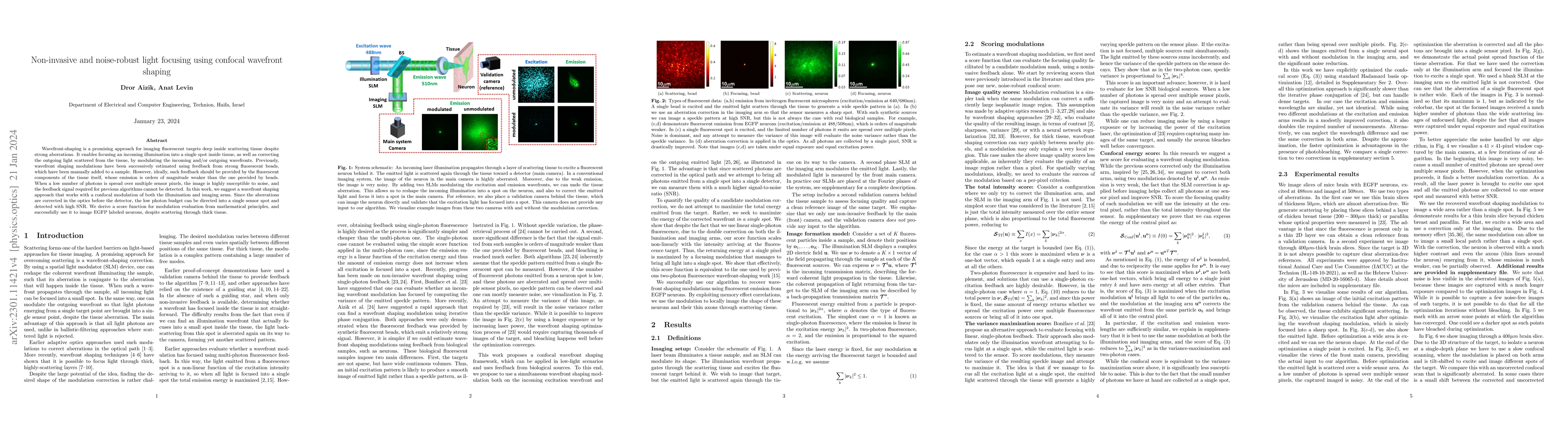
Wavefront-shaping is a promising approach for imaging fluorescent targets deep inside scattering tissue despite strong aberrations. It enables focusing an incoming illumination into a single spot inside tissue, as well as correcting the outgoing light scattered from the tissue, by modulating the incoming and/or outgoing wavefronts. Previously, wavefront shaping modulations have been successively estimated using feedback from strong fluorescent beads, which have been manually added to a sample. However, ideally, such feedback should be provided by the fluorescent components of the tissue itself, whose emission is orders of magnitude weaker than the one provided by beads. When a low number of photons is spread over multiple sensor pixels, the image is highly susceptible to noise, and the feedback signal required for previous algorithms cannot be detected. In this work, we suggest a wavefront shaping approach that works with a confocal modulation of both the illumination and imaging arms. Since the aberrations are corrected in the optics before the detector, the low photon budget can be directed into a single sensor spot and detected with high SNR. We derive a score function for modulation evaluation from mathematical principles, and successfully use it to image EGFP labeled neurons, despite scattering through thick tissue.
AI Key Findings
Get AI-generated insights about this paper's methodology, results, significance, and more — seven facets brought into focus.
Impact
Paper Details
Authors
PDF Preview
Key Terms
Citation Network
Current paper (gray), citations (green), references (blue)
Display is limited for performance on very large graphs.
Discussion 0