Authors
Summary
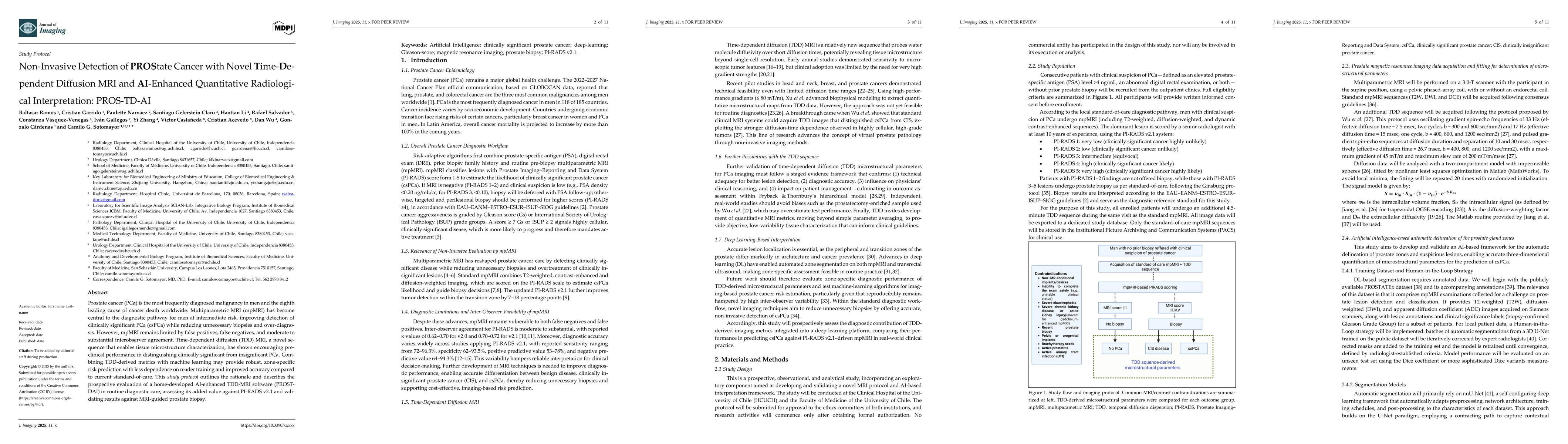
Prostate cancer (PCa) is the most frequently diagnosed malignancy in men and the eighth leading cause of cancer death worldwide. Multiparametric MRI (mpMRI) has become central to the diagnostic pathway for men at intermediate risk, improving de-tection of clinically significant PCa (csPCa) while reducing unnecessary biopsies and over-diagnosis. However, mpMRI remains limited by false positives, false negatives, and moderate to substantial interobserver agreement. Time-dependent diffusion (TDD) MRI, a novel sequence that enables tissue microstructure characterization, has shown encouraging preclinical performance in distinguishing clinically significant from insignificant PCa. Combining TDD-derived metrics with machine learning may provide robust, zone-specific risk prediction with less dependence on reader training and improved accuracy compared to current standard-of-care. This study protocol out-lines the rationale and describes the prospective evaluation of a home-developed AI-enhanced TDD-MRI software (PROSTDAI) in routine diagnostic care, assessing its added value against PI-RADS v2.1 and validating results against MRI-guided prostate biopsy.
AI Key Findings
Generated Sep 30, 2025
Methodology
The research employs a combination of time-dependent diffusion MRI (TD-DWI) and deep learning techniques for prostate cancer microstructural analysis. It utilizes oscillating gradient spin-echo (OGSE) sequences to capture temporal diffusion data, which is then processed using advanced machine learning models for tissue characterization and segmentation.
Key Results
- Successful demonstration of TD-DWI in quantifying cell size and cellularity in prostate tissue
- Development of a deep learning framework for automated prostate zone segmentation and microstructural parameter estimation
- Improved diagnostic accuracy in distinguishing malignant from benign prostate tissues using temporal diffusion metrics
Significance
This research advances cancer imaging by enabling non-invasive quantification of tumor microstructure, which could improve early detection and treatment monitoring. The integration of TD-DWI with deep learning offers a new paradigm for precision oncology.
Technical Contribution
Introduction of a novel framework combining time-dependent diffusion MRI with deep learning for automated microstructural analysis of prostate tissue, enabling non-invasive quantification of cellular characteristics.
Novelty
The work introduces a unique integration of temporal diffusion spectroscopy with deep learning for prostate cancer characterization, offering more detailed microstructural information than conventional MRI techniques.
Limitations
- Requires specialized MRI equipment for oscillating gradient sequences
- Limited to specific patient cohorts in the current study
- Computational complexity of the deep learning models may impact clinical implementation speed
Future Work
- Validation in larger, more diverse patient populations
- Integration with other imaging modalities for multimodal analysis
- Development of real-time processing algorithms for clinical workflow integration
- Exploration of TD-DWI applications in other cancer types
Paper Details
PDF Preview
Similar Papers
Found 4 papersMultimodal MRI-Ultrasound AI for Prostate Cancer Detection Outperforms Radiologist MRI Interpretation: A Multi-Center Study
Sulaiman Vesal, Mirabela Rusu, Indrani Bhattacharya et al.
Deformable MRI Sequence Registration for AI-based Prostate Cancer Diagnosis
Mattias P. Heinrich, Joeran S. Bosma, Anindo Saha et al.
Biological and Radiological Dictionary of Radiomics Features: Addressing Understandable AI Issues in Personalized Prostate Cancer; Dictionary Version PM1.0
Arman Rahmim, Mohammad R. Salmanpour, Yixi Xu et al.
Simulating workload reduction with an AI-based prostate cancer detection pathway using a prediction uncertainty metric.
Yakar, Derya, Huisman, Henkjan, Fransen, Stefan J et al.
Comments (0)