Publication
Metrics
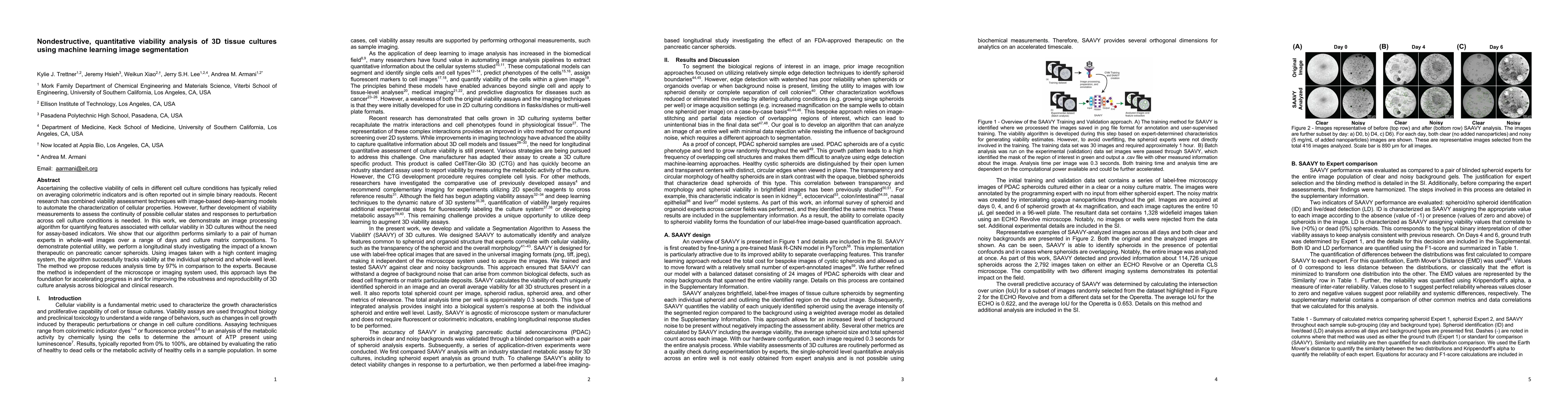
AI Quick Summary
This paper presents a machine learning-based image segmentation algorithm for nondestructive, quantitative viability analysis of 3D tissue cultures, achieving results comparable to human experts while significantly reducing analysis time by 97%. The method successfully tracks viability changes in pancreatic cancer spheroids over time, demonstrating its potential to enhance 3D culture analysis in biological and clinical research.
Paper Preview
Abstract
Ascertaining the collective viability of cells in different cell culture conditions has typically relied on averaging colorimetric indicators and is often reported out in simple binary readouts. Recent research has combined viability assessment techniques with image-based deep-learning models to automate the characterization of cellular properties. However, further development of viability measurements to assess the continuity of possible cellular states and responses to perturbation across cell culture conditions is needed. In this work, we demonstrate an image processing algorithm for quantifying cellular viability in 3D cultures without the need for assay-based indicators. We show that our algorithm performs similarly to a pair of human experts in whole-well images over a range of days and culture matrix compositions. To demonstrate potential utility, we perform a longitudinal study investigating the impact of a known therapeutic on pancreatic cancer spheroids. Using images taken with a high content imaging system, the algorithm successfully tracks viability at the individual spheroid and whole-well level. The method we propose reduces analysis time by 97% in comparison to the experts. Because the method is independent of the microscope or imaging system used, this approach lays the foundation for accelerating progress in and for improving the robustness and reproducibility of 3D culture analysis across biological and clinical research.
AI Key Findings
Get AI-generated insights about this paper's methodology, results, significance, and more — seven facets brought into focus.
Impact
Paper Details
Authors
PDF Preview
Key Terms
Citation Network
Current paper (gray), citations (green), references (blue)
Display is limited for performance on very large graphs.
Discussion 0