Quick Summary:

This paper proposes a novel STU-Net approach using transfer learning to enhance brain tissue segmentation in nonhuman primates (NHPs) by leveraging human brain MRI data. The method achieves high accuracy in segmenting small subcortical structures, demonstrating significant improvements in Dice Similarity Coefficient, Intersection over Union, and Hausdorff Distance 95, potentially advancing research in evolutionary neuroscience and neurological disorders.

Summary

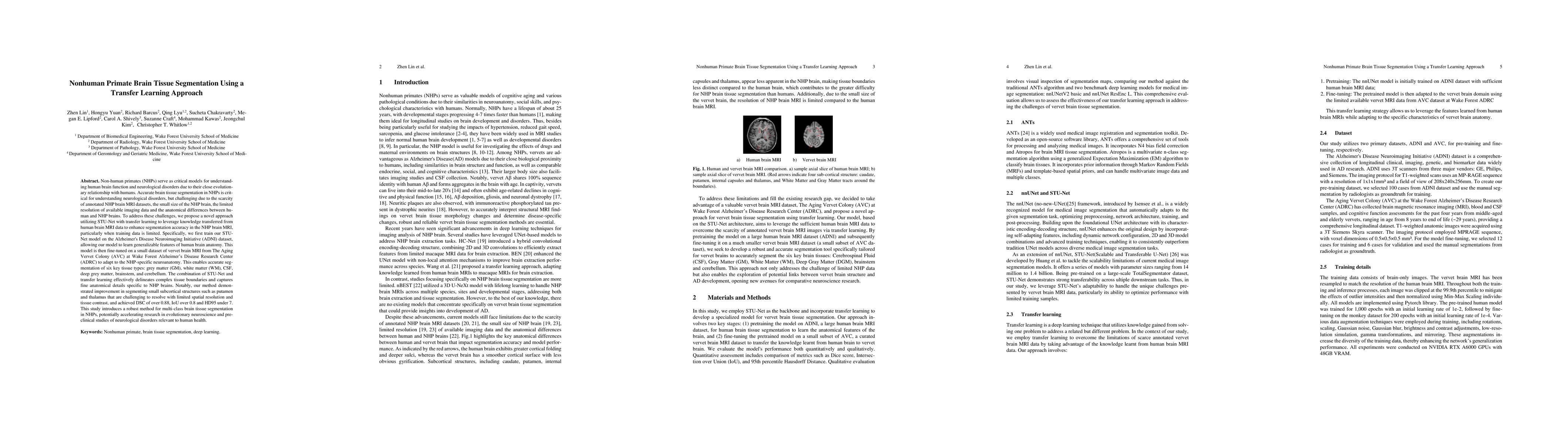
Non-human primates (NHPs) serve as critical models for understanding human brain function and neurological disorders due to their close evolutionary relationship with humans. Accurate brain tissue segmentation in NHPs is critical for understanding neurological disorders, but challenging due to the scarcity of annotated NHP brain MRI datasets, the small size of the NHP brain, the limited resolution of available imaging data and the anatomical differences between human and NHP brains. To address these challenges, we propose a novel approach utilizing STU-Net with transfer learning to leverage knowledge transferred from human brain MRI data to enhance segmentation accuracy in the NHP brain MRI, particularly when training data is limited. The combination of STU-Net and transfer learning effectively delineates complex tissue boundaries and captures fine anatomical details specific to NHP brains. Notably, our method demonstrated improvement in segmenting small subcortical structures such as putamen and thalamus that are challenging to resolve with limited spatial resolution and tissue contrast, and achieved DSC of over 0.88, IoU over 0.8 and HD95 under 7. This study introduces a robust method for multi-class brain tissue segmentation in NHPs, potentially accelerating research in evolutionary neuroscience and preclinical studies of neurological disorders relevant to human health.

AI Key Findings

Generated Jun 10, 2025

Methodology

The research proposes a novel approach using STU-Net with transfer learning to segment nonhuman primate (NHP) brain tissue, addressing challenges like limited annotated NHP MRI datasets, small brain size, low resolution, and anatomical differences from human brains. The method involves pretraining on human brain MRI data and fine-tuning on limited vervet MRI data.

Key Results

  • The proposed method outperforms traditional ANTs and two deep learning benchmarks (nnUNetV2 basic and nnUNet ResEnc L) in Dice Score, IoU, and 95th percentile Hausdorff Distance across six brain tissue types.
  • The model excels in boundary delineation, reducing 95th percentile Hausdorff Distance by half compared to ANTs and showing significant improvements over the other deep learning benchmarks.
  • The study demonstrates robustness and accuracy in capturing fine anatomical details and boundaries, particularly in challenging sub-cortical regions.

Significance

This research introduces a robust method for multi-class brain tissue segmentation in NHPs, potentially accelerating research in evolutionary neuroscience and preclinical studies of neurological disorders relevant to human health.

Technical Contribution

The paper presents a transfer learning-based approach using STU-Net for NHP brain tissue segmentation, demonstrating improved performance over conventional methods and other deep learning benchmarks.

Novelty

The research combines STU-Net with transfer learning to address the scarcity of annotated NHP brain MRI data, effectively adapting human-trained models to NHP brain segmentation, particularly for small subcortical structures.

Limitations

  • The training dataset was relatively small, which may limit the model's ability to generalize to diverse NHP populations.
  • The study was conducted on a single NHP species, restricting its applicability to other species with different brain morphologies.
  • Reliance on T1-weighted MRI data alone may not fully capture tissue contrasts relevant for precise segmentation.

Future Work

  • Incorporate larger, more diverse datasets and multimodal MRI sequences to address current limitations.
  • Explore the integration of T2-weighted and diffusion MRI to enhance tissue contrast and segmentation accuracy.
  • Expand the approach to multiple NHP species to improve model generalizability across different anatomical structures.

Paper Details

PDF Preview

Download PDF

Key Terms

No key terms available for this paper.

No citations found for this paper.

Comments (0)

This website uses cookies to ensure you get the best experience. Learn more