Summary
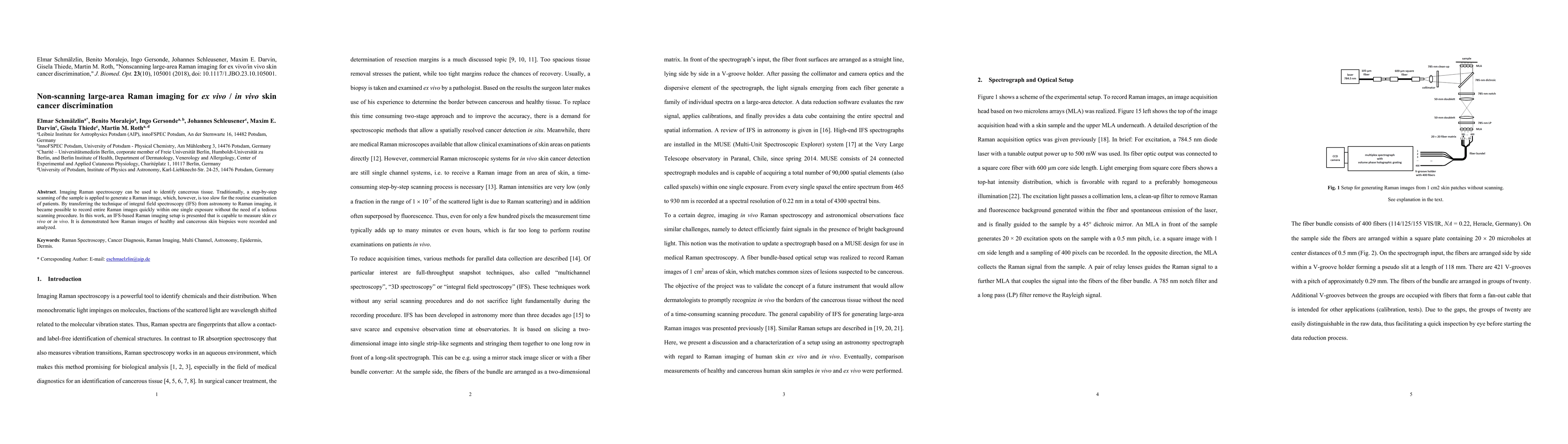
Imaging Raman spectroscopy can be used to identify cancerous tissue. Traditionally, a step-by-step scanning of the sample is applied to generate a Raman image, which, however, is too slow for routine examination of patients. By transferring the technique of integral field spectroscopy (IFS) from astronomy to Raman imaging, it becomes possible to record entire Raman images quickly within a single exposure, without the need for a tedious scanning procedure. An IFS-based Raman imaging setup is presented, which is capable of measuring skin ex vivo or in vivo. It is demonstrated how Raman images of healthy and cancerous skin biopsies were recorded and analyzed.
AI Key Findings
Get AI-generated insights about this paper's methodology, results, and significance.
Paper Details
PDF Preview
Key Terms
Citation Network
Current paper (gray), citations (green), references (blue)
Display is limited for performance on very large graphs.
Similar Papers
Found 4 papersLiving human lung slices for ex vivo modelling of lung cancer.
Winter, Hauke, Schneider, Marc A, Savai, Rajkumar et al.
| Title | Authors | Year | Actions |
|---|
Comments (0)