Quick Summary:

This study evaluates the normalized blood flow index (NBFI) from optical coherence tomography angiography (OCTA) as a biomarker for early diabetic retinopathy (DR). The NBFI in the deep capillary plexus (DCP) was found to be the most sensitive indicator, distinguishing early DR from healthy controls better than blood vessel density and blood flow flux.

Summary

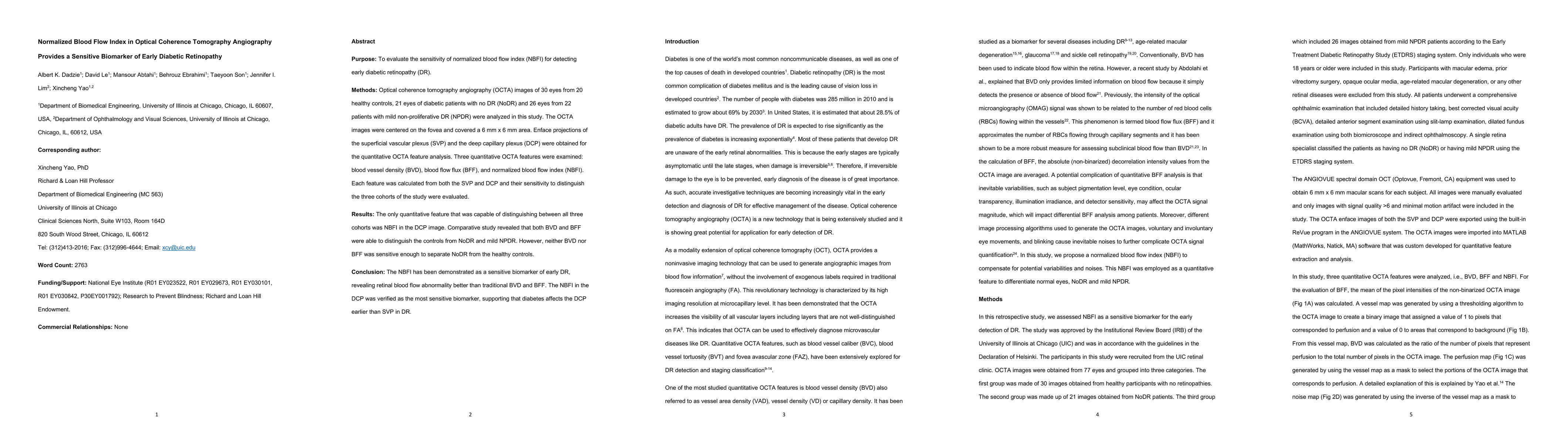
Purpose: To evaluate the sensitivity of normalized blood flow index (NBFI) for detecting early diabetic retinopathy (DR). Methods: Optical coherence tomography angiography (OCTA) images of 30 eyes from 20 healthy controls, 21 eyes of diabetic patients with no DR (NoDR) and 26 eyes from 22 patients with mild non-proliferative DR (NPDR) were analyzed in this study. The OCTA images were centered on the fovea and covered a 6 mm x 6 mm area. Enface projections of the superficial vascular plexus (SVP) and the deep capillary plexus (DCP) were obtained for the quantitative OCTA feature analysis. Three quantitative OCTA features were examined: blood vessel density (BVD), blood flow flux (BFF), and normalized blood flow index (NBFI). Each feature was calculated from both the SVP and DCP and their sensitivity to distinguish the three cohorts of the study were evaluated. Results: The only quantitative feature that was capable of distinguishing between all three cohorts was NBFI in the DCP image. Comparative study revealed that both BVD and BFF were able to distinguish the controls from NoDR and mild NPDR. However, neither BVD nor BFF was sensitive enough to separate NoDR from the healthy controls. Conclusion: The NBFI has been demonstrated as a sensitive biomarker of early DR, revealing retinal blood flow abnormality better than traditional BVD and BFF. The NBFI in the DCP was verified as the most sensitive biomarker, supporting that diabetes affects the DCP earlier than SVP in DR.

AI Key Findings

Get AI-generated insights about this paper's methodology, results, and significance.

Paper Details

PDF Preview

Download PDF

Key Terms

dr (0.355)
controls (0.261)
controls (0.259)
healthy (0.244)
quantitative (0.214)
flow (0.213)
mm (0.210)
quantitative (0.209)
mild (0.209)
patients (0.209)
patients (0.209)
normalized (0.206)
mild (0.205)
sensitive (0.205)
distinguish (0.200)
mm (0.200)
flow (0.199)
sensitive (0.196)
distinguish (0.186)
normalized (0.184)

Citation Network

Current paper (gray), citations (green), references (blue)

Current Paper
Citations
References
Click to view
Title Authors Year Actions
Differential artery-vein analysis in OCTA for predicting the anti-VEGF treatment outcome of diabetic macular edema
Mansour Abtahi, Albert K Dadzie, B. Ebrahimi, Boda Huang, Yi-Ting Hsieh, Xincheng Yao
2025
View Paper
Early Diabetic Retinopathy Evaluation With OCTA: A Study on Vascular Branching and Fragmentation.
Yao Yu, Shiwei Cui, Yang He, Jiahao Zhang, Nan Lu, Yanqiu Yang, Jian Liu, Yi Wang, Zhenhe Ma
2024
View Paper
Freqformer: Frequency-Domain Transformer for 3-D Visualization and Quantification of Human Retinal Circulation
Lingyun Wang, Bingjie Wang, Jay Chhablani, J. Sahel, Shaohua Pi
2024
View Paper
OCTA-ReVA: an open-source toolbox for comprehensive retinal vessel feature analysis in optical coherence tomography angiography
Albert K Dadzie, David Le, Mansour Abtahi, B. Ebrahimi, Tobiloba Adejumo, Taeyoon Son, Michael J. Heiferman, Jennifer I. Lim, Xincheng Yao
2024
View Paper
Differential Capillary and Large Vessel Analysis Improves OCTA Classification of Diabetic Retinopathy
Mansour Abtahi, David Le, B. Ebrahimi, Albert K Dadzie, Mojtaba Rahimi, Yi-Ting Hsieh, Michael J. Heiferman, Jennifer I. Lim, Xincheng Yao
2024
View Paper
Potential Diagnostic Biomarkers of tRNA-Derived Small RNAs in PBMCs for Nonproliferative Diabetic Retinopathy in Patients With Type 2 Diabetes Mellitus
Chun Ding, Nan Wang, Aohua Peng, Zicong Wang, Bingyan Li, Xian Zhang, Jun Zeng, Yedi Zhou
2024
View Paper
Differential artery-vein analysis improves the OCTA classification of diabetic retinopathy
Mansour Abtahi, David Le, B. Ebrahimi, Albert K Dadzie, Mojtaba Rahimi, Yi-Ting Hsieh, Michael J. Heiferman, Jennifer I. Lim, Xincheng Yao
2024
View Paper
OCT angiography 2023 update: focus on diabetic retinopathy.
Emanuele Crincoli, R. Sacconi, L. Querques, Giuseppe Querques
2024
View Paper
Deep learning-based optical coherence tomography angiography image construction using spatial vascular connectivity network
David Le, Taeyoon Son, Tae-Hoon Kim, Tobiloba Adejumo, Mansour Abtahi, Shaiban Ahmed, Alfa Rossi, B. Ebrahimi, Albert K Dadzie, Guangying Ma, Jennifer I. Lim, Xincheng Yao
2024
View Paper
Adaptive vessel tracing and segmentation in OCT enables the robust detection of wall-to-lumen ratio abnormalities in 5xFAD mice
Tobiloba Adejumo, Guangying Ma, Taeyoon Son, Tae-Hoon Kim, David Le, Albert K Dadzie, Shaiban Ahmed, Xincheng Yao
2023
View Paper
Deep learning for artery–vein classification in optical coherence tomography angiography
David Le, Mansour Abtahi, Tobiloba Adejumo, B. Ebrahimi, Albert K Dadzie, Taeyoon Son, Xincheng Yao
2023
View Paper

Comments (0)

This website uses cookies to ensure you get the best experience. Learn more