Normative brain mapping using scalp EEG and potential clinical application
Publication
Metrics
AI Quick Summary
This study constructs stable normative brain maps of scalp EEG relative band power from healthy controls, correlating them with MEG and intracranial EEG. The findings suggest these maps could be useful for lateralizing abnormal regions in epilepsy, indicating potential clinical applications.
Paper Preview
Abstract
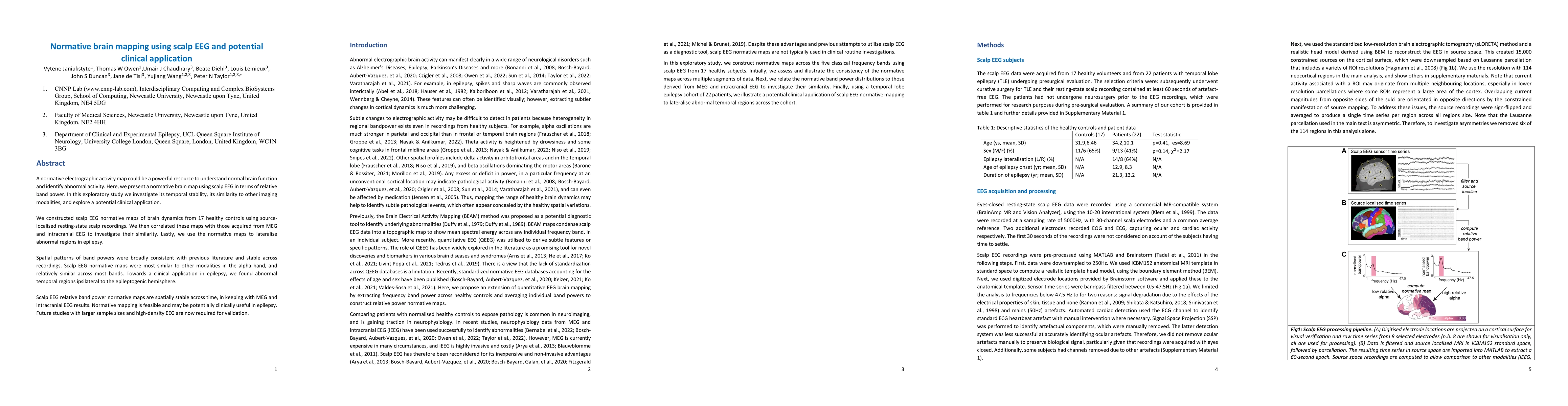
A normative electrographic activity map could be a powerful resource to understand normal brain function and identify abnormal activity. Here, we present a normative brain map using scalp EEG in terms of relative band power. In this exploratory study we investigate its temporal stability, its similarity to other imaging modalities, and explore a potential clinical application. We constructed scalp EEG normative maps of brain dynamics from 17 healthy controls using source-localised resting-state scalp recordings. We then correlated these maps with those acquired from MEG and intracranial EEG to investigate their similarity. Lastly, we use the normative maps to lateralise abnormal regions in epilepsy. Spatial patterns of band powers were broadly consistent with previous literature and stable across recordings. Scalp EEG normative maps were most similar to other modalities in the alpha band, and relatively similar across most bands. Towards a clinical application in epilepsy, we found abnormal temporal regions ipsilateral to the epileptogenic hemisphere. Scalp EEG relative band power normative maps are spatially stable across time, in keeping with MEG and intracranial EEG results. Normative mapping is feasible and may be potentially clinically useful in epilepsy. Future studies with larger sample sizes and high-density EEG are now required for validation.
AI Key Findings
Get AI-generated insights about this paper's methodology, results, significance, and more — seven facets brought into focus.
Impact
Paper Details
Authors
PDF Preview
Key Terms
Citation Network
Current paper (gray), citations (green), references (blue)
Display is limited for performance on very large graphs.
Discussion 0