NucleiMix is a data augmentation method designed to address data imbalance in nuclei instance segmentation by increasing the representation of rare nuclei types. It combines candidate location identification and a diffusion model-based inpainting strategy to realistically integrate rare nuclei, improving segmentation and classification performance.
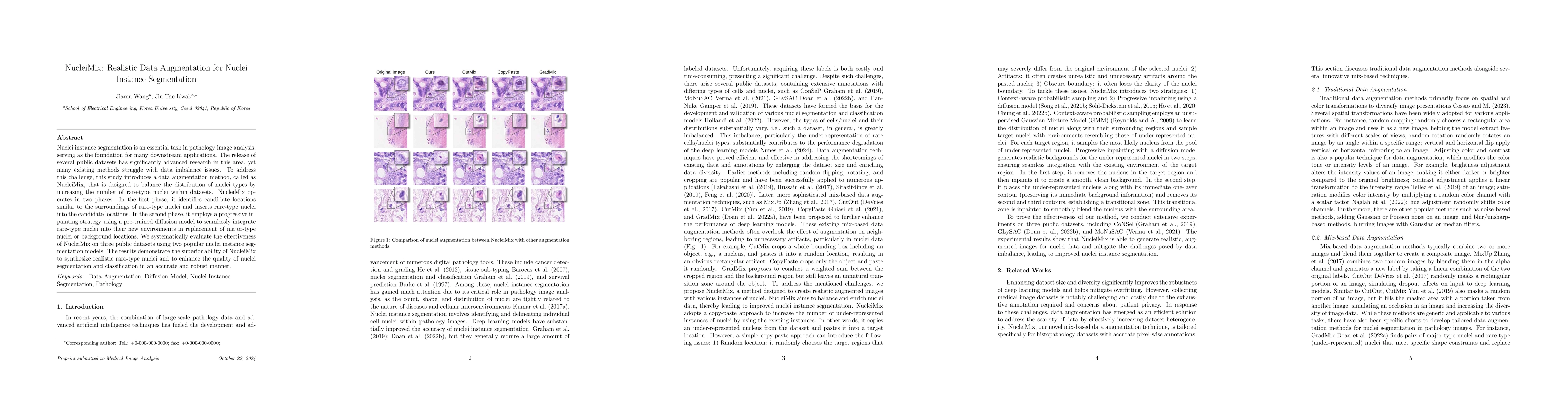
Nuclei instance segmentation is an essential task in pathology image analysis, serving as the foundation for many downstream applications. The release of several public datasets has significantly advanced research in this area, yet many existing methods struggle with data imbalance issues. To address this challenge, this study introduces a data augmentation method, called NucleiMix, which is designed to balance the distribution of nuclei types by increasing the number of rare-type nuclei within datasets. NucleiMix operates in two phases. In the first phase, it identifies candidate locations similar to the surroundings of rare-type nuclei and inserts rare-type nuclei into the candidate locations. In the second phase, it employs a progressive inpainting strategy using a pre-trained diffusion model to seamlessly integrate rare-type nuclei into their new environments in replacement of major-type nuclei or background locations. We systematically evaluate the effectiveness of NucleiMix on three public datasets using two popular nuclei instance segmentation models. The results demonstrate the superior ability of NucleiMix to synthesize realistic rare-type nuclei and to enhance the quality of nuclei segmentation and classification in an accurate and robust manner.
Get AI-generated insights about this paper's methodology, results, and significance.
Current paper (gray), citations (green), references (blue)
No similar papers found for this research.
No citations found for this paper.
Title | Authors | Year | Actions |
---|---|---|---|
A Survey on Cell Nuclei Instance Segmentation and Classification: Leveraging Context and Attention
|
João D. Nunes, D. Montezuma, Domingos Oliveira, Tania Pereira, Jaime S. Cardoso
|
2024
|
View Paper |
Augmenting Medical Imaging: A Comprehensive Catalogue of 65 Techniques for Enhanced Data Analysis
|
M. Cossio
|
2023
|
View Paper |
Diffusion Posterior Sampling for General Noisy Inverse Problems
|
Hyungjin Chung, Jeongsol Kim, Michael T. McCann, M. Klasky, J. C. Ye
|
2022
|
View Paper |
Conditional GANs based system for fibrosis detection and quantification in Hematoxylin and Eosin whole slide images
|
A. Naglah, F. Khalifa, A. El-Baz, Dibson D. Gondim
|
2022
|
View Paper |
Transformer-based unsupervised contrastive learning for histopathological image classification
|
Xiyue Wang, Sen Yang, Jun Zhang, Minghui Wang, Jing Zhang, Wei Yang, Junzhou Huang, Xiao Han
|
2022
|
View Paper |
SONNET: A Self-Guided Ordinal Regression Neural Network for Segmentation and Classification of Nuclei in Large-Scale Multi-Tissue Histology Images
|
Tan N. N. Doan, Boram Song, T. Vuong, Kyungeun Kim, J. T. Kwak
|
2022
|
View Paper |
Nucleus segmentation: towards automated solutions.
|
Réka Hollandi, Nikita Moshkov, L. Paavolainen, Ervin A. Tasnádi, F. Piccinini, P. Horváth
|
2022
|
View Paper |
Generative Adversarial Networks
|
I. Goodfellow, Jean Pouget-Abadie, Mehdi Mirza, Bing Xu, David Warde-Farley, Sherjil Ozair, Aaron C. Courville, Yoshua Bengio
|
2021
|
View Paper |
PointNu-Net: Keypoint-Assisted Convolutional Neural Network for Simultaneous Multi-Tissue Histology Nuclei Segmentation and Classification
|
Kai Yao, Kaizhu Huang, Jie Sun, Amir Hussain
|
2021
|
View Paper |
MoNuSAC2020: A Multi-Organ Nuclei Segmentation and Classification Challenge
|
R. Verma, Neeraj Kumar, Abhijeet Patil, N. Kurian, S. Rane, S. Graham, Q. Vu, Mieke Zwager, S. Raza, N. Rajpoot, Xiyi Wu, Huai Chen, Yijie Huang, Lisheng Wang, Hyun Jung, G. Brown, Yanling Liu, Shuolin Liu, Seyed Abolghassem Fatemi Jahromi, A. Khani, Ehsan Montahaei, M. Baghshah, Hamid Behroozi, P. Semkin, Alexandr G. Rassadin, Prasad Dutande, Romil Lodaya, U. Baid, B. Baheti, S. Talbar, A. Mahbod, R. Ecker, I. Ellinger, Zhipeng Luo, Bin Dong, Zhengyu Xu, Yuehan Yao, Shuai Lv, Ming Feng, Kele Xu, H. Zunair, A. Hamza, S. Smiley, T. Yin, Qianhao Fang, S. Srivastava, D. Mahapatra, Lubomira Trnavska, Hanyun Zhang, P. Narayanan, Justin Law, Yinyin Yuan, Abhiroop Tejomay, Aditya Mitkari, D. Koka, V. Ramachandra, L. Kini, A. Sethi
|
2021
|
View Paper |
Diffusion Models Beat GANs on Image Synthesis
|
Prafulla Dhariwal, Alex Nichol
|
2021
|
View Paper |
Simple Copy-Paste is a Strong Data Augmentation Method for Instance Segmentation
|
Golnaz Ghiasi, Yin Cui, A. Srinivas, Rui Qian, Tsung-Yi Lin, E. D. Cubuk, Quoc V. Le, Barret Zoph
|
2020
|
View Paper |
Score-Based Generative Modeling through Stochastic Differential Equations
|
Yang Song, Jascha Narain Sohl-Dickstein, Diederik P. Kingma, Abhishek Kumar, Stefano Ermon, Ben Poole
|
2020
|
View Paper |
CutMix: Regularization Strategy to Train Strong Classifiers With Localizable Features
|
Sangdoo Yun, Dongyoon Han, Seong Joon Oh, Sanghyuk Chun, Junsuk Choe, Y. Yoo
|
2019
|
View Paper |
PanNuke: An Open Pan-Cancer Histology Dataset for Nuclei Instance Segmentation and Classification
|
Jevgenij Gamper, Navid Alemi Koohbanani, Ksenija Benet, A. Khuram, N. Rajpoot
|
2019
|
View Paper |
Data Augmentation for Chest Pathologies Classification
|
I. Sirazitdinov, Maksym Kholiavchenko, R. Kuleev, B. Ibragimov
|
2019
|
View Paper |
Quantifying the effects of data augmentation and stain color normalization in convolutional neural networks for computational pathology
|
David Tellez, G. Litjens, Péter Bándi, W. Bulten, J. Bokhorst, F. Ciompi, J. Laak
|
2019
|
View Paper |
Hover-Net: Simultaneous segmentation and classification of nuclei in multi-tissue histology images
|
S. Graham, Q. Vu, S. Raza, A. Azam, Y. Tsang, J. T. Kwak, N. Rajpoot
|
2018
|
View Paper |
Data Augmentation Using Random Image Cropping and Patching for Deep CNNs
|
Ryo Takahashi, Takashi Matsubara, K. Uehara
|
2018
|
View Paper |
Methods for Segmentation and Classification of Digital Microscopy Tissue Images
|
Q. Vu, S. Graham, Minh Nguyen Nhat To, M. Shaban, Talha Qaiser, Navid Alemi Koohbanani, S. Khurram, T. Kurç, K. Farahani, Tianhao Zhao, Rajarsi R. Gupta, J. T. Kwak, N. Rajpoot, J. Saltz
|
2018
|
View Paper |
Gaussian Mixture Models
|
D. Reynolds
|
2018
|
View Paper |
Panoptic Segmentation
|
Alexander Kirillov, Kaiming He, Ross B. Girshick, C. Rother, Piotr Dollár
|
2018
|
View Paper |
mixup: Beyond Empirical Risk Minimization
|
Hongyi Zhang, Moustapha Cissé, Yann Dauphin, David Lopez-Paz
|
2017
|
View Paper |
Improved Regularization of Convolutional Neural Networks with Cutout
|
Terrance Devries, Graham W. Taylor
|
2017
|
View Paper |
A Dataset and a Technique for Generalized Nuclear Segmentation for Computational Pathology
|
Neeraj Kumar, R. Verma, Sanuj Sharma, S. Bhargava, Abhishek Vahadane, A. Sethi
|
2017
|
View Paper |
Deep Unsupervised Learning using Nonequilibrium Thermodynamics
|
Jascha Narain Sohl-Dickstein, Eric A. Weiss, Niru Maheswaranathan, S. Ganguli
|
2015
|
View Paper |
Histology image analysis for carcinoma detection and grading
|
Lei He, L. Long, Sameer Kiran Antani, G. Thoma
|
2012
|
View Paper |
Tweedie’s Formula and Selection Bias
|
B. Efron
|
2011
|
View Paper |
The Navier–Stokes–Voight model for image inpainting
|
M. Ebrahimi, M. Holst, E. Lunasin
|
2009
|
View Paper |
Information Retrieval
|
Kareem Darwish
|
2008
|
View Paper |
Renal cell carcinoma sub‐typing by histopathology and fluorescence in situ hybridization on a needle‐biopsy specimen
|
D. Barocas, S. Mathew, J. DelPizzo, E. Vaughan, R. Sosa, R. Fine, M. Akhtar, D. Scherr
|
2007
|
View Paper |
Artificial neural networks improve the accuracy of cancer survival prediction
|
H. Burke, P. Goodman, D. B. Rosen, D. Henson, J. Weinstein, Frank E. Harrell, J. Marks, D. Winchester, D. Bostwick
|
1997
|
View Paper |
”a deep learning approach for colonoscopy pathology wsi analysis: accurate segmentation and classi fi cation”
|
2020
|
||
”denoising di ff usion probabilistic models”
|
2020
|
||
”di ff erential data augmentation techniques for medical imaging classi fi cation tasks”
|
2017
|
||
”reverse-time di ff usion equation models”
|
1982
|
||
2022b. ”improving di ff usion models for inverse problems using manifold constraints”
|
Showing first page preview. Click "Open Full PDF" to view the complete paper.
Your comment contains inappropriate language and has been rejected.
Are you sure you want to remove this tag? This action cannot be undone.
Someone just favorited a paper!
Comments (0)