Nuquantus: Machine learning software for the characterization and quantification of cell nuclei in complex immunofluorescent tissue images
Publication
Metrics
Paper Preview
Abstract
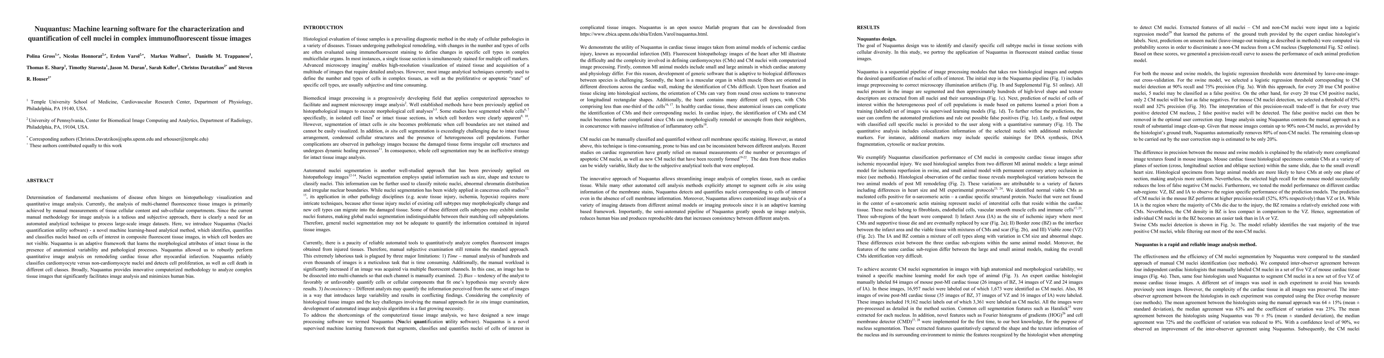
Determination of fundamental mechanisms of disease often hinges on histopathology visualization and quantitative image analysis. Currently, the analysis of multi-channel fluorescence tissue images is primarily achieved by manual measurements of tissue cellular content and sub-cellular compartments. Since the current manual methodology for image analysis is a tedious and subjective approach, there is clearly a need for an automated analytical technique to process large-scale image datasets. Here, we introduce Nuquantus (Nuclei quantification utility software) - a novel machine learning-based analytical method, which identifies, quantifies and classifies nuclei based on cells of interest in composite fluorescent tissue images, in which cell borders are not visible. Nuquantus is an adaptive framework that learns the morphological attributes of intact tissue in the presence of anatomical variability and pathological processes. Nuquantus allowed us to robustly perform quantitative image analysis on remodeling cardiac tissue after myocardial infarction. Nuquantus reliably classifies cardiomyocyte versus non-cardiomyocyte nuclei and detects cell proliferation, as well as cell death in different cell classes. Broadly, Nuquantus provides innovative computerized methodology to analyze complex tissue images that significantly facilitates image analysis and minimizes human bias.
AI Key Findings
Get AI-generated insights about this paper's methodology, results, significance, and more — seven facets brought into focus.
Impact
Paper Details
PDF Preview
Key Terms
Citation Network
Current paper (gray), citations (green), references (blue)
Display is limited for performance on very large graphs.
Discussion 0