Publication
Metrics
AI Quick Summary
OCTAVA is an open-source toolbox designed to standardize the quantitative analysis of optical coherence tomography angiography (OCTA) images, facilitating automated pre-processing, segmentation, and metric determination for microvasculature characterization across different instruments. Its wide adoption could enable large-scale data aggregation to develop reliable biomarkers for early detection and treatment of microvascular diseases.
Paper Preview
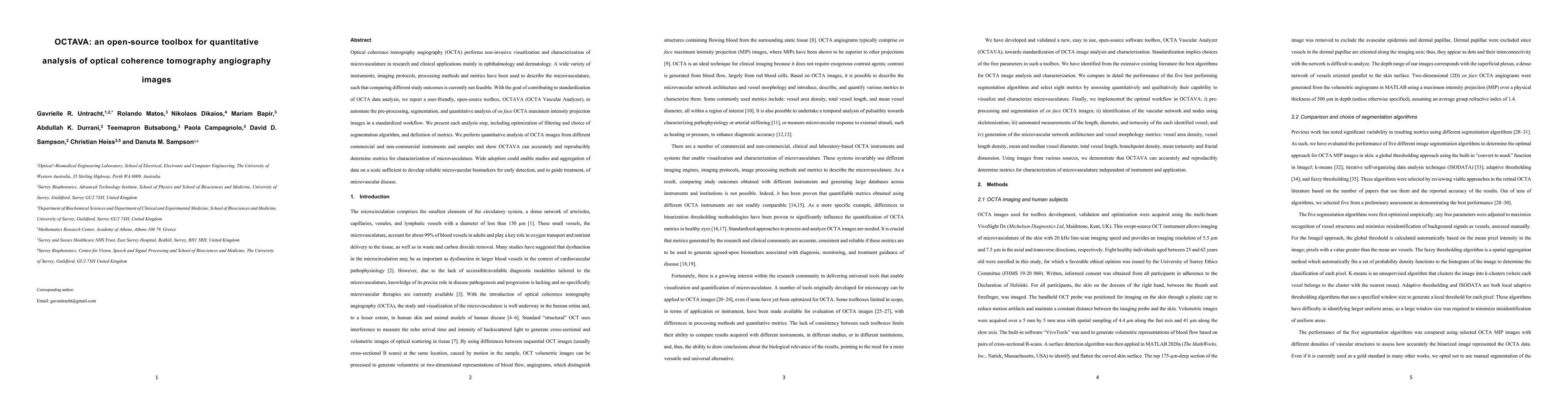
Abstract
Optical coherence tomography angiography (OCTA) performs non-invasive visualization and characterization of microvasculature in research and clinical applications mainly in ophthalmology and dermatology. A wide variety of instruments, imaging protocols, processing methods and metrics have been used to describe the microvasculature, such that comparing different study outcomes is currently not feasible. With the goal of contributing to standardization of OCTA data analysis, we report a user-friendly, open-source toolbox, OCTAVA (OCTA Vascular Analyzer), to automate the pre-processing, segmentation, and quantitative analysis of en face OCTA maximum intensity projection images in a standardized workflow. We present each analysis step, including optimization of filtering and choice of segmentation algorithm, and definition of metrics. We perform quantitative analysis of OCTA images from different commercial and non-commercial instruments and samples and show OCTAVA can accurately and reproducibly determine metrics for characterization of microvasculature. Wide adoption could enable studies and aggregation of data on a scale sufficient to develop reliable microvascular biomarkers for early detection, and to guide treatment, of microvascular disease.
AI Key Findings
Get AI-generated insights about this paper's methodology, results, significance, and more — seven facets brought into focus.
Impact
Paper Details
Authors
PDF Preview
Key Terms
Citation Network
Current paper (gray), citations (green), references (blue)
Display is limited for performance on very large graphs.
Discussion 0