01
MethodologyHow they did it
The OCTID database was created using a raster scan protocol with a 2mm scan length and 512x1024 pixel resolution.
The OCTID database was created using a raster scan protocol with a 2mm scan length and 512x1024 pixel resolution. More in Methodology →
The database contains over 500 high-resolution images categorized into different pathological conditions. — 25 normal OCT images were included with their corresponding ground truth delineations for accurate evaluation of OCT image segmentation. More in Key Results →
OCTID provides a comprehensive open-access database for clinical ophthalmology, enabling early diagnosis of retinal diseases. More in Significance →
The dataset may not be representative of all retinal conditions or patient populations. — Ground truth delineations were limited to 25 normal images. More in Limitations →
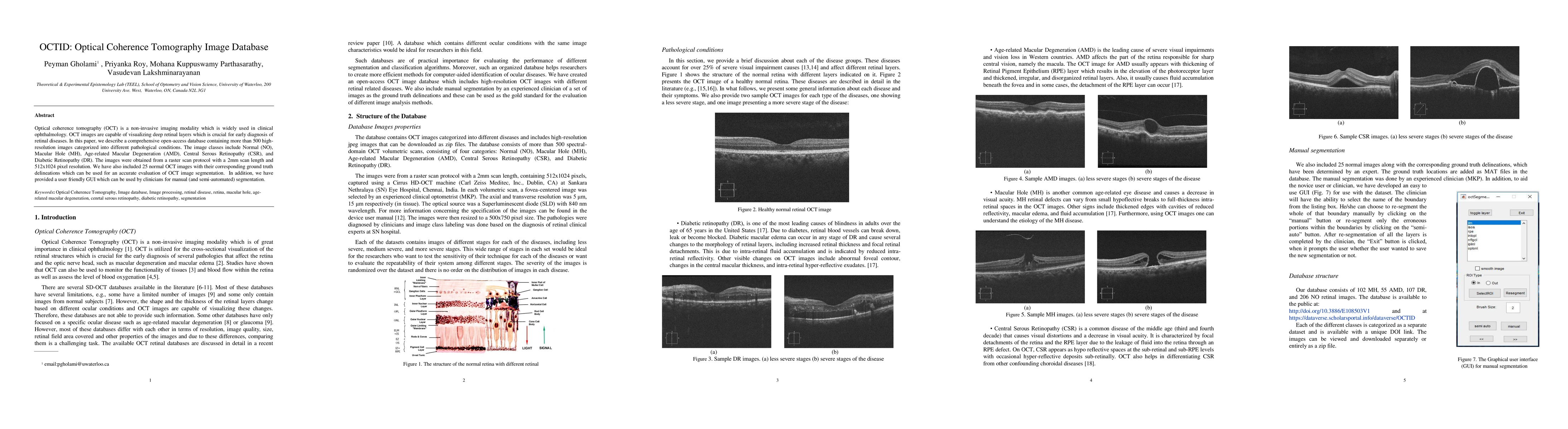
Optical coherence tomography (OCT) is a non-invasive imaging modality which is widely used in clinical ophthalmology. OCT images are capable of visualizing deep retinal layers which is crucial for early diagnosis of retinal diseases. In this paper, we describe a comprehensive open-access database containing more than 500 highresolution images categorized into different pathological conditions. The image classes include Normal (NO), Macular Hole (MH), Age-related Macular Degeneration (AMD), Central Serous Retinopathy (CSR), and Diabetic Retinopathy (DR). The images were obtained from a raster scan protocol with a 2mm scan length and 512x1024 pixel resolution. We have also included 25 normal OCT images with their corresponding ground truth delineations which can be used for an accurate evaluation of OCT image segmentation. In addition, we have provided a user-friendly GUI which can be used by clinicians for manual (and semi-automated) segmentation.
Seven facets of this paper, analysed and brought into focus by AI.
OCTID provides a comprehensive open-access database for clinical ophthalmology, enabling early diagnosis of retinal diseases.
The OCTID database was created using a raster scan protocol with a 2mm scan length and 512x1024 pixel resolution.
OCTID provides a comprehensive open-access database for clinical ophthalmology, enabling early diagnosis of retinal diseases.
The creation of a comprehensive open-access OCT image database provides a valuable resource for researchers and clinicians.
OCTID is the first publicly available database of high-resolution OCT images, enabling large-scale studies and collaborative research.
Current paper (gray), citations (green), references (blue)
Display is limited for performance on very large graphs.
Discussion 0