This paper proposes SEENet, a semi-automatic framework for one-click lesion segmentation and RECIST measurement on CT scans, significantly reducing radiologists' workload and improving measurement reliability. The framework refines initial lesion estimates with minimal user input, achieving state-of-the-art performance on the DeepLesion dataset.
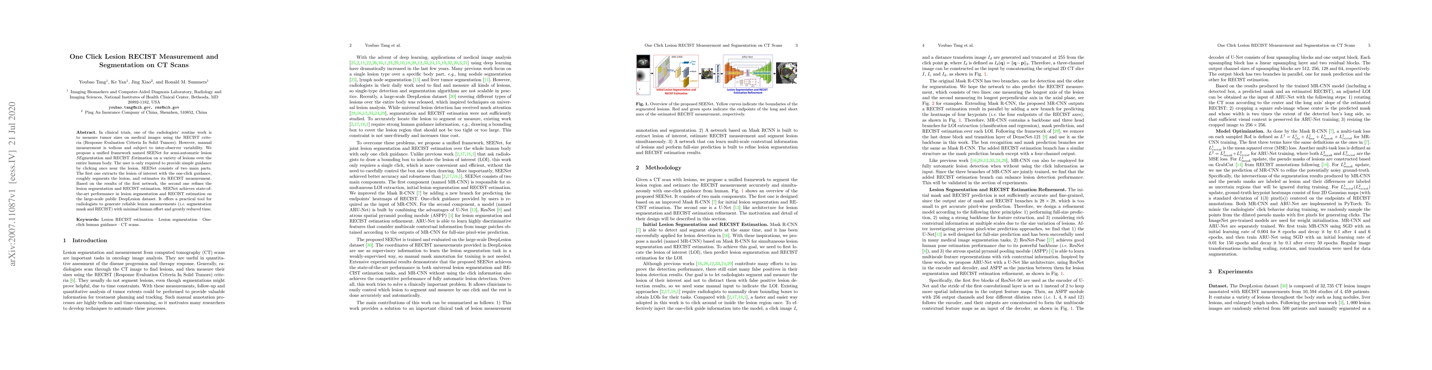
In clinical trials, one of the radiologists' routine work is to measure tumor sizes on medical images using the RECIST criteria (Response Evaluation Criteria In Solid Tumors). However, manual measurement is tedious and subject to inter-observer variability. We propose a unified framework named SEENet for semi-automatic lesion \textit{SE}gmentation and RECIST \textit{E}stimation on a variety of lesions over the entire human body. The user is only required to provide simple guidance by clicking once near the lesion. SEENet consists of two main parts. The first one extracts the lesion of interest with the one-click guidance, roughly segments the lesion, and estimates its RECIST measurement. Based on the results of the first network, the second one refines the lesion segmentation and RECIST estimation. SEENet achieves state-of-the-art performance in lesion segmentation and RECIST estimation on the large-scale public DeepLesion dataset. It offers a practical tool for radiologists to generate reliable lesion measurements (i.e. segmentation mask and RECIST) with minimal human effort and greatly reduced time.
This graph shows how this paper is connected to other research. The current paper is shown in dark gray, papers that cite it are in green, and papers it references are in blue. You can:
• Hover over any node to see the paper title
• Click a node to view that paper's details
• Drag nodes to rearrange the graph
• Use the zoom controls (bottom right) to zoom in/out
Title | Authors | Year | Actions |
---|---|---|---|
Dedicated software to harmonize the follow-up of oncological patients
|
Mathias Illy, Axel Bartoli, Julien Mancini, Florence Duffaud, Vincent Vidal, F. Tradi
|
2024
|
View Paper |
The ULS23 Challenge: a Baseline Model and Benchmark Dataset for 3D Universal Lesion Segmentation in Computed Tomography
|
M.J.J. de Grauw, E.Th. Scholten, E. Smit, M.J.C.M. Rutten, M. Prokop, B. V. Ginneken, A. Hering
|
2024
|
View Paper |
Improving assessment of lesions in longitudinal CT scans: a bi-institutional reader study on an AI-assisted registration and volumetric segmentation workflow
|
Alessa Hering, Max Westphal, Annika Gerken, Haidara Almansour, Michael Maurer, Benjamin Geisler, Temke Kohlbrandt, Thomas Eigentler, Teresa Amaral, N. Lessmann, S. Gatidis, Horst K. Hahn, Konstantin Nikolaou, Ahmed E. Othman, J. Moltz, Felix Peisen
|
2024
|
View Paper |
Optimizing Universal Lesion Segmentation: State Space Model-Guided Hierarchical Networks with Feature Importance Adjustment
|
Kazi Shahriar Sanjid, Md. Tanzim Hossain, Md. Shakib Shahariar Junayed, M. M. Uddin
|
2024
|
View Paper |
Large-scale long-tailed disease diagnosis on radiology images
|
Qiaoyu Zheng, Weike Zhao, Chaoyi Wu, Xiaoman Zhang, Ya Zhang, Yanfeng Wang, Weidi Xie
|
2023
|
View Paper |
Deep Interactive Segmentation of Medical Images: A Systematic Review and Taxonomy
|
Zdravko Marinov, Paul F. Jäger, Jan Egger, J. Kleesiek, Rainer Stiefelhagen
|
2023
|
View Paper |
Scale-aware Test-time Click Adaptation for Pulmonary Nodule and Mass Segmentation
|
Zhihao Li, Jiancheng Yang, Yongchao Xu, Li Zhang, Wen-Wen Dong, Bo Du
|
2023
|
View Paper |
RECIST-Induced Reliable Learning: Geometry-Driven Label Propagation for Universal Lesion Segmentation
|
Lianyu Zhou, Lequan Yu, Liansheng Wang
|
2023
|
View Paper |
RECIST Weakly Supervised Lesion Segmentation via Label-Space Co-Training
|
Lianyu Zhou, Dong Wei, Donghuan Lu, Wei Xue, Liansheng Wang, Yefeng Zheng
|
2023
|
View Paper |
Accurate and Robust Lesion RECIST Diameter Prediction and Segmentation with Transformers
|
Youbao Tang, Ning Zhang, Yirui Wang, Shenghua He, Mei Han, Jing Xiao, Ruei-Sung Lin
|
2022
|
View Paper |
ImplicitAtlas: Learning Deformable Shape Templates in Medical Imaging
|
Jiancheng Yang, Udaranga Wickramasinghe, Bingbing Ni, P. Fua
|
2022
|
View Paper |
AGMR-Net: Attention-guided multiscale recovery framework for stroke segmentation
|
Xiuquan Du, Kunpeng Ma, Yuhui Song
|
2022
|
View Paper |
RECISTSup: Weakly-Supervised Lesion Volume Segmentation Using RECIST Measurement
|
Han Wang, Fasheng Yi, Jingling Wang, Zhang Yi, Haixian Zhang
|
2022
|
View Paper |
Twenty Years On: RECIST as a Biomarker of Response in Solid Tumours an EORTC Imaging Group – ESOI Joint Paper
|
L. Fournier, L. de Geus-Oei, D. Regge, D. Oprea-Lager, M. D’Anastasi, L. Bidaut, T. Bäuerle, E. Lopci, G. Cappello, F. Lecouvet, M. Mayerhoefer, W. Kunz, J. Verhoeff, D. Caruso, M. Smits, R. Hoffmann, S. Gourtsoyianni, R. Beets-Tan, E. Neri, N. deSouza, C. Deroose, C. Caramella
|
2022
|
View Paper |
Lesion Segmentation and RECIST Diameter Prediction via Click-driven Attention and Dual-path Connection
|
Youbao Tang, K. Yan, Jinzheng Cai, Lingyun Huang, G. Xie, Jing Xiao, Jingjing Lu, Gigin Lin, Le Lu
|
2021
|
View Paper |
Weakly-Supervised Universal Lesion Segmentation with Regional Level Set Loss
|
Youbao Tang, Jinzheng Cai, K. Yan, Lingyun Huang, G. Xie, Jing Xiao, Jingjing Lu, Gigin Lin, Le Lu
|
2021
|
View Paper |
A 2021 update on cancer image analytics with deep learning
|
N. Kurian, A. Sethi, Anil Reddy Konduru, A. Mahajan, S. Rane
|
2021
|
View Paper |
Deep Lesion Tracker: Monitoring Lesions in 4D Longitudinal Imaging Studies
|
Jinzheng Cai, Youbao Tang, K. Yan, Adam P. Harrison, Jing Xiao, Gigin Lin, Le Lu
|
2020
|
View Paper |
A Review of Deep Learning in Medical Imaging: Imaging Traits, Technology Trends, Case Studies With Progress Highlights, and Future Promises
|
S. K. Zhou, H. Greenspan, C. Davatzikos, J. Duncan, B. Ginneken, A. Madabhushi, Jerry L Prince, D. Rueckert, R. Summers
|
2020
|
View Paper |
E2Net: An Edge Enhanced Network for Accurate Liver and Tumor Segmentation on CT Scans
|
Youbao Tang, Yuxing Tang, Yingying Zhu, Jing Xiao, R. Summers
|
2020
|
View Paper |
Comments (0)