OpenWSI is a low-cost, high-throughput whole slide imaging system utilizing single-frame autofocusing and open-source hardware components, enabling comparable throughput to high-end platforms while significantly reducing costs. The system employs a programmable LED array, CNC router, and ultrasonic motor for precise axial positioning, achieving a resolution of ~0.7 microns.
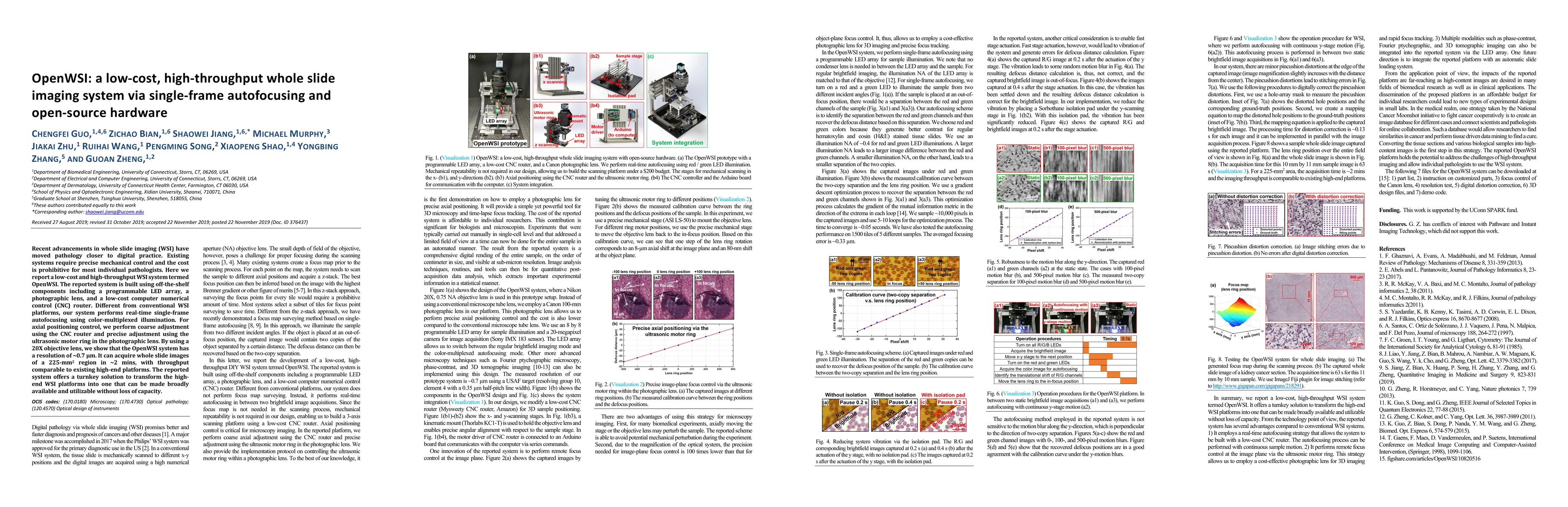
Recent advancements in whole slide imaging (WSI) have moved pathology closer to digital practice. Existing systems require precise mechanical control and the cost is prohibitive for most individual pathologists. Here we report a low-cost and high-throughput WSI system termed OpenWSI. The reported system is built using off-the-shelf components including a programmable LED array, a photographic lens, and a low-cost computer numerical control (CNC) router. Different from conventional WSI platforms, our system performs real-time single-frame autofocusing using color-multiplexed illumination. For axial positioning control, we perform coarse adjustment using the CNC router and precise adjustment using the ultrasonic motor ring in the photographic lens. By using a 20X objective lens, we show that the OpenWSI system has a resolution of ~0.7 microns. It can acquire whole slide images of a 225-mm^2 region in ~2 mins, with throughput comparable to existing high-end platforms. The reported system offers a turnkey solution to transform the high-end WSI platforms into one that can be made broadly available and utilizable without loss of capacity.
Get AI-generated insights about this paper's methodology, results, and significance.
Current paper (gray), citations (green), references (blue)
Title | Authors | Year | Actions |
---|---|---|---|
SparseFocus: Learning-based One-shot Autofocus for Microscopy with Sparse Content
|
Yongping Zhai, Xiaoxi Fu, Qiang Su, Jia Hu, Yake Zhang, Yunfeng Zhou, Chaofan Zhang, Xiao Li, Wenxin Wang, Dongdong Wu, Shen Yan
|
2025
|
View Paper |
Rapid Autofocus Method Based on LED Oblique Illumination for Metaphase Chromosome Microscopy Imaging System
|
Changliang Yu, Fangqiu Ding, Zhenyu Ma, Yuguo Tang
|
2024
|
View Paper |
Cost-effective scanning digital microscopic system for wide field-of-view and high-resolution biomedical imaging
|
Siru Lin, Zhihang Lv, Xiwei Huang
|
2024
|
View Paper |
Efficient, gigapixel-scale, aberration-free whole slide scanner using angular ptychographic imaging with closed-form solution
|
Shi Zhao, Haowen Zhou, S. Lin, Ruizhi Cao, Changhuei Yang
|
2024
|
View Paper |
High-resolution display screen as programmable illumination for Fourier ptychography
|
K. Lee, K. Lee, Jaewoo Jung, Hyesuk Chae, Seung Ah Lee
|
2024
|
View Paper |
White blood cells segmentation and classification using a random forest and residual networks implementation
|
Marlon R. Rodrigues Garcia, Erika Toneth Ponce Ayala, S. Pratavieira, Vanderlei Salvador Bagnato
|
2024
|
View Paper |
Analytical linear model for Fourier ptychography: insights into image uniqueness and reconstruction factors
|
Pengcheng Lou, S. Veetil, Xiaoliang He, Aihui Sun, Shouyu Wang, Cheng Liu
|
2024
|
View Paper |
Fourier Ptychographic Microscopy 10 Years on: A Review
|
Fannuo Xu, Zipei Wu, Chao Tan, Yizheng Liao, Zhiping Wang, Keru Chen, An Pan
|
2024
|
View Paper |
Low-cost, autonomous microscopy using deep learning and robotics: A crystal morphology case study
|
Matthew R. Wilkinson, B. Castro-Dominguez, Chick C. Wilson, Uriel Martinez-Hernandez
|
2023
|
View Paper |
Large-scale lensless microscopy with fast acquisition and region-wise focusing
|
You Zhou, Weizhi Song, Linyuan Wu, Lin Fan, Junjia Wang, Shaowei Jiang, Zhan Ma, Bo Xiong, Xun Cao
|
2023
|
View Paper |
Lensless polarimetric coded ptychography for high-resolution, high-throughput gigapixel birefringence imaging on a chip
|
Liming Yang, Ruihai Wang, Qianhao Zhao, Pengming Song, Shaowei Jiang, Tianbo Wang, Xiaopeng Shao, Chengfei Guo, Rishikesh Pandey, G. Zheng
|
2023
|
View Paper |
A review on low‐cost microscopes for Open Science
|
J. Salido, G. Bueno, Jesús Ruiz-Santaquiteria, G. Cristóbal
|
2022
|
View Paper |
Analysis of postreconstruction digital refocusing in Fourier ptychographic microscopy
|
Haowen Zhou, Cheng Shen, Mingshu Liang, Changhuei Yang
|
2022
|
View Paper |
Automated Open-Hardware Multiwell Imaging Station for Microorganisms Observation
|
Alain Gervasi, P. Cardol, Patrick E. Meyer
|
2022
|
View Paper |
Fourier Ptychographic Microscopy via Alternating Direction Method of Multipliers
|
Aiye Wang, Zhuoqun Zhang, Siqi Wang, An Pan, Caiwen Ma, Baoli Yao
|
2022
|
View Paper |
Resolution doubling in optofluidics and sample-scanning fluorescence microscopy
|
B. Mandracchia, Kidan Tadesse, Jeonghwan Son, Shu Jia
|
2022
|
View Paper |
Regression Based Iterative Illumination Compensation Method for Multi-Focal Whole Slide Imaging System
|
Róbert Paulik, M. Kozlovszky, B. Molnár
|
2021
|
View Paper |
Resolution-Enhanced Parallel Coded Ptychography for High-Throughput Optical Imaging
|
Shaowei Jiang, Chengfei Guo, Pengming Song, Niyun Zhou, Z. Bian, Jiakai Zhu, Ruihai Wang, Pei Dong, Zibang Zhang, Jun Liao, Jianhua Yao, B. Feng, Michael Murphy, G. Zheng
|
2021
|
View Paper |
Ptychographic sensor for large-scale lensless microbial monitoring with high spatiotemporal resolution.
|
Shaowei Jiang, Chengfei Guo, Z. Bian, Ruihai Wang, Jiakai Zhu, Pengming Song, Patrick Hu, Derek Hu, Zibang Zhang, K. Hoshino, Bin Feng, G. Zheng
|
2021
|
View Paper |
High-throughput lensless whole slide imaging via continuous height-varying modulation of a tilted sensor.
|
Shaowei Jiang, Chengfei Guo, P. Hu, Derek Hu, Pengming Song, Tianbo Wang, Z. Bian, Zibang Zhang, G. Zheng
|
2021
|
View Paper |
Low-cost whole slide imaging system with single-shot autofocusing based on color-multiplexed illumination and deep learning.
|
Kaifa Xin, Shaowei Jiang, Xu Chen, Yonghong He, Jian Zhang, Hongpeng Wang, Honghai Liu, Qin Peng, Yongbing Zhang, Xiangyang Ji
|
2021
|
View Paper |
Recent technical advances in whole slide imaging instrumentation
|
Prateek Katare, S. S. Gorthi
|
2021
|
View Paper |
Single-Shot Through-Focus Image Acquisition and Phase Retrieval From Chromatic Aberration and Multi-Angle Illumination
|
Guocheng Zhou, Shaohui Zhang, Yayu Zhai, Yao Hu, Q. Hao
|
2021
|
View Paper |
Fast digital refocusing and depth of field extended Fourier ptychography microscopy.
|
Shaohui Zhang, Guocheng Zhou, Chuan-wen Zheng, Tong Li, Yao Hu, Q. Hao
|
2021
|
View Paper |
High throughput slanted scanning whole slide imaging system for digital pathology
|
Ning Li, Tao Lv, Yulin Sun, Xiuli Liu, Shaoqun Zeng, Xiaohua Lv
|
2021
|
View Paper |
Concept, implementations and applications of Fourier ptychography
|
G. Zheng, Cheng Shen, Shaowei Jiang, Pengming Song, Changhuei Yang
|
2021
|
View Paper |
Concept, implementations and applications of Fourier ptychography
|
G. Zheng, Cheng Shen, Shaowei Jiang, Pengming Song, Changhuei Yang
|
2021
|
View Paper |
High-throughput fast full-color digital pathology based on Fourier ptychographic microscopy via color transfer
|
Yuting Gao, Jiurun Chen, Ai-hong Wang, An Pan, Caiwen Ma, Baoli Yao
|
2021
|
View Paper |
Contemporary Whole Slide Imaging Devices and Their Applications within the Modern Pathology Department: A Selected Hardware Review
|
Ankush U Patel, Ulysses Balis, Jerome Cheng, Zaibo Li, Giovanni M. Lujan, D. McClintock, L. Pantanowitz, A. Parwani
|
2021
|
View Paper |
Squid: Simplifying Quantitative Imaging Platform Development and Deployment
|
Hongquan Li, Deepak Krishnamurthy, Ethan Li, P. Vyas, Nibha Akireddy, Chew Chai, Manu Prakash
|
2020
|
View Paper |
Speeded-Up Robust Features-based image mosaic method for large-scale microscopic hyperspectral pathological imaging
|
Qing Zhang, Li Sun, Jiangang Chen, Mei Zhou, Menghan Hu, Ying Wen, Qingli Li
|
2020
|
View Paper |
Progress and prospect of biomedical optical imaging
|
L. Fu, Qingming Luo
|
2020
|
View Paper |
Edge effect removal in Fourier ptychographic microscopy via perfect Fourier transformation (PFT)
|
An Pan, Junfu Zheng, Chuansheng Xia, Yuting Gao, Caiwen Ma, Baoli Yao
|
2020
|
View Paper |
Edge effect removal in Fourier ptychographic microscopy via periodic plus smooth image decomposition
|
An Pan, Aiye Wang, Junfu Zheng, Yuting Gao, Caiwen Ma, Baoli Yao
|
2020
|
View Paper |
Virtual brightfield and fluorescence staining for Fourier ptychography via unsupervised deep learning.
|
Ruihai Wang, Pengming Song, Shaowei Jiang, C. Yan, Jiakai Zhu, Chengfei Guo, Z. Bian, Tianbo Wang, G. Zheng
|
2020
|
View Paper |
Coherent synthetic aperture imaging for visible remote sensing via reflective Fourier ptychography.
|
Meng Xiang, An Pan, Yi-yi Zhao, XueWu Fan, Hui Zhao, Chuang Li, Baoli Yao
|
2020
|
View Paper |
High-resolution and large field-of-view Fourier ptychographic microscopy and its applications in biomedicine
|
An Pan, Chao Zuo, Baoli Yao
|
2020
|
View Paper |
Autofocusing technologies for whole slide imaging and automated microscopy
|
Z. Bian, Chengfei Guo, Shaowei Jiang, Jiakai Zhu, Ruihai Wang, Pengming Song, Zibang Zhang, K. Hoshino, G. Zheng
|
2020
|
View Paper |
Simple Shading Correction Method for Brightfield Whole Slide Imaging
|
Yoon-Oh Tak, Anjin Park, Jang-Un Choi, J. Eom, H. Kwon, J. Eom
|
2020
|
View Paper |
Whole Slide Imaging (WSI) in Pathology: Current Perspectives and Future Directions
|
Neeta Kumar, Ruchika Gupta, Sanjay Gupta
|
2020
|
View Paper |
无透镜编码叠层显微成像原理及研究进展(特邀)
|
郭成飞 Guo Chengfei, 李婧妍 Li Jingyan, 江劭玮 Jiang Shaowei, 邵晓鹏 Shao Xiaopeng, 郑国安 Zheng Guoan
|
2024
|
View Paper |
Real-Time, Automated, Multiobjective, Cloud Computing Whole Slide Imaging Device
|
S. Mallick, K. S. Adithya, R. Sabui, Balu Babu Naidu, Ranjeet Singh, H. Babu, Gajula Jagadeesh, K. M. Venkata Amaresh, R. Gopal, V. Sharma
|
2023
|
View Paper |
基于傅里叶叠层显微成像术的定量相干传递函数恢复
|
陈佳铭 CHEN Jiaming, 潘安 Pan An, 王爱业 Wang Aiye, 马彩文 Ma Caiwen, 姚保利 Yao Baoli
|
2023
|
View Paper |
Brightfield, fluorescence, and phase-contrast whole slide imaging via dual-LED autofocusing
|
UO Chengfeig, Z. I. B. Ian, S. O. A. Lhudaithy, S. H. J. Iang, Y. U. T. Omizawa, P. E. S. Ong, T. I. W. Ang, A. X. I. S. Hao
|
2021
|
View Paper |
Quantitative Retrieval of Coherent Transfer Function via Fourier Ptychographic Microscopy
|
View Paper |
Comments (0)