Publication
Metrics
AI Quick Summary
This paper proposes a learning-based method to estimate the boundary of objects in electromagnetic medical imaging using the same imaging data, avoiding additional sensors and movement issues. The method utilizes reflection coefficients and is validated through clinical trials, achieving an average dissimilarity of 0.012 in Hu-moment for head boundary detection.
Paper Preview
Abstract
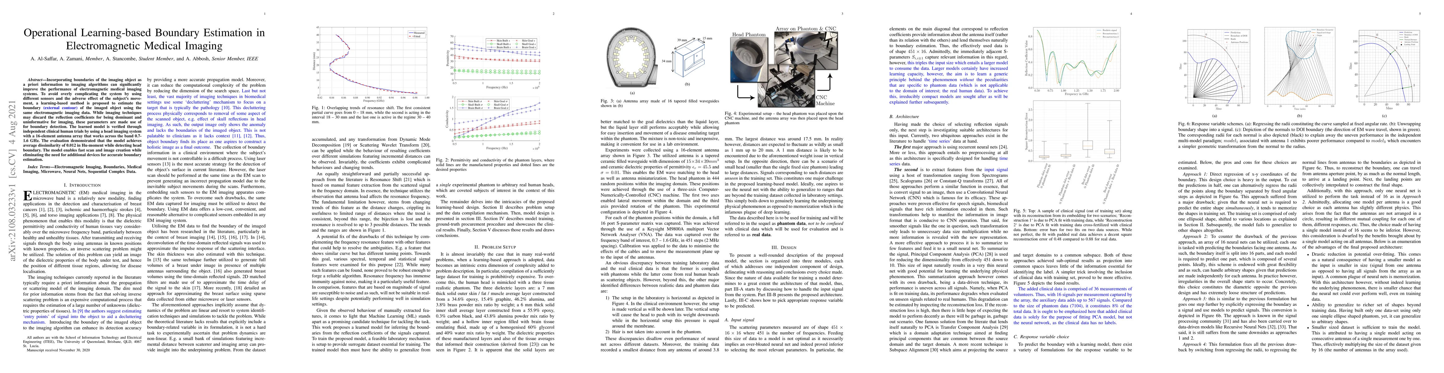
Incorporating boundaries of the imaging object as a priori information to imaging algorithms can significantly improve the performance of electromagnetic medical imaging systems. To avoid overly complicating the system by using different sensors and the adverse effect of the subject's movement, a learning-based method is proposed to estimate the boundary (external contour) of the imaged object using the same electromagnetic imaging data. While imaging techniques may discard the reflection coefficients for being dominant and uninformative for imaging, these parameters are made use of for boundary detection. The learned model is verified through independent clinical human trials by using a head imaging system with a 16-element antenna array that works across the band 0.7-1.6 GHz. The evaluation demonstrated that the model achieves average dissimilarity of 0.012 in Hu-moment while detecting head boundary. The model enables fast scan and image creation while eliminating the need for additional devices for accurate boundary estimation.
AI Key Findings
Get AI-generated insights about this paper's methodology, results, significance, and more — seven facets brought into focus.
Impact
Paper Details
Authors
PDF Preview
Key Terms
Citation Network
Current paper (gray), citations (green), references (blue)
Display is limited for performance on very large graphs.
Discussion 0