This paper provides a tutorial on optimizing image capture for low-light widefield quantitative fluorescence microscopy, focusing on the use of specialised cameras like EMCCD, qCMOS, and sCMOS to minimise illumination and reduce photobleaching and phototoxicity in live biological samples. It discusses performance factors, noise sources, user-controllable parameters, and post-processing algorithms to enhance image quality for quantitative measurements.
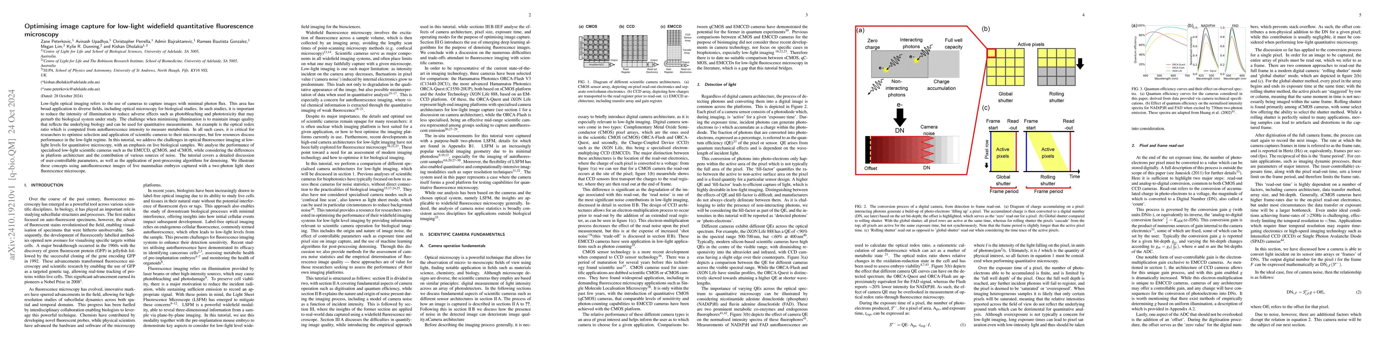
Low-light optical imaging refers to the use of cameras to capture images with minimal photon flux. This area has broad application to diverse fields, including optical microscopy for biological studies. In such studies, it is important to reduce the intensity of illumination to reduce adverse effects such as photobleaching and phototoxicity that may perturb the biological system under study. The challenge when minimising illumination is to maintain image quality that reflects the underlying biology and can be used for quantitative measurements. An example is the optical redox ratio which is computed from autofluorescence intensity to measure metabolism. In all such cases, it is critical for researchers to optimise selection and application of scientific cameras to their microscopes, but few resources discuss performance in the low-light regime. In this tutorial, we address the challenges in optical fluorescence imaging at low-light levels for quantitative microscopy, with an emphasis on live biological samples. We analyse the performance of specialised low-light scientific cameras such as the EMCCD, qCMOS, and sCMOS, while considering the differences in platform architecture and the contribution of various sources of noise. The tutorial covers a detailed discussion of user-controllable parameters, as well as the application of post-processing algorithms for denoising. We illustrate these concepts using autofluorescence images of live mammalian embryos captured with a two-photon light sheet fluorescence microscope.
Get AI-generated insights about this paper's methodology, results, and significance.
Current paper (gray), citations (green), references (blue)
Priya Singh, J. -P. Tetienne, A. J. Healey et al.
Apratim Majumder, Rajesh Menon, Richard G. Baird
Title | Authors | Year | Actions |
---|---|---|---|
A neuromorphic camera for tracking passive and active matter with lower data throughput
|
Gabriel Britto Monteiro, Megan Lim, T. Tan, Avinash Upadhya, Zhuo Liang, Benjamin Agnew, Tomonori Hu, Benjamin J. Eggleton, Chris Perrella, Kylie R. Dunning, Kishan Dholakia
|
2025
|
View Paper |
Title | Authors | Year | Actions |
---|---|---|---|
Quantifying DNA damage following light sheet and confocal imaging of the mammalian embryo
|
D. Chow, E. Schartner, Stella Corsetti, Avinash Upadhya, Josephine M. Morizet, F. Gunn-Moore, Kylie R. Dunning, Kishan Dholakia
|
2024
|
View Paper |
A comparison between the measurement of quantum spatial correlations using qCMOS photon-number resolving and electron multiplying CCD camera technologies
|
K. Roberts, O. Wolley, T. Gregory, M. Padgett
|
2024
|
View Paper |
Molecular histopathology of matrix proteins through autofluorescence super-resolution microscopy
|
Biswajoy Ghosh, J. Chatterjee, R. Paul, Sebastián Acuña, P. Lahiri, M. Pal, Pabitra Mitra, Krishna Agarwal
|
2024
|
View Paper |
Viewing early life without labels: optical approaches for imaging the early embryo
|
D. Chow, T. Tan, Avinash Upadhya, M. Lim, Kishan Dholakia, K. Dunning
|
2024
|
View Paper |
High sensitivity cameras can lower spatial resolution in high-resolution optical microscopy
|
Henning Ortkrass, Marcel Müller, Anders Kokkvoll Engdahl, Gerhard Holst, Thomas Huser
|
2024
|
View Paper |
Metabolic imaging of human embryos is predictive of ploidy status but is not associated with clinical pregnancy outcomes: a pilot trial.
|
D. Sakkas, C. Gulliford, G. Ardestani, O. Ocali, Marion Martins, Nitya Talasila, Jaimin S. Shah, A. Penzias, E. Seidler, T. Sanchez
|
2024
|
View Paper |
Restoration of metabolic functional metrics from label-free, two-photon human tissue images using multiscale deep-learning-based denoising algorithms
|
Nilay Vora, C. Polleys, Filippos Sakellariou, Georgios Georgalis, Hong-Thao Thieu, Elisabeth Genega, Narges Jahanseir, Abani Patra, Eric Miller, Irene Georgakoudi
|
2023
|
View Paper |
Label-free biomedical optical imaging
|
N. Shaked, S. Boppart, Lihong V. Wang, J. Popp
|
2023
|
View Paper |
Choosing the Best Camera System for Your Biological Light Microscopy Needs: Part II
|
W. G. (. Jerome
|
2023
|
View Paper |
UVA Hyperspectral Light-Sheet Microscopy for Volumetric Metabolic Imaging: Application to Preimplantation Embryo Development
|
Josephine M. Morizet, D. Chow, Philip Wijesinghe, E. Schartner, George O. Dwapanyin, Nicolas S. Dubost, G. Bruce, Ellen Anckaert, Kylie R. Dunning, Kishan Dholakia
|
2023
|
View Paper |
Light-sheet autofluorescence lifetime imaging with a single-photon avalanche diode array
|
K. Samimi, D. Desa, W. Lin, K. Weiss, Joe Li, J. Huisken, V. Miskolci, A. Huttenlocher, J. Chacko, A. Velten, J. Rogers, K. Eliceiri, M. Skala
|
2023
|
View Paper |
Viewing life without labels under optical microscopes
|
Biswajoy Ghosh, Krishna Agarwal
|
2023
|
View Paper |
Light sheet autofluorescence lifetime imaging with a single photon avalanche diode array
|
K. Samimi, D. Desa, Wei Lin, K. Weiss, Joe Li, J. Huisken, V. Miskolci, A. Huttenlocher, J. Chacko, A. Velten, J. Rogers, K. Eliceiri, M. Skala
|
2023
|
View Paper |
Practical considerations for quantitative light sheet fluorescence microscopy
|
C. Hobson, Min Guo, Harshad D. Vishwasrao, Yicong Wu, H. Shroff, T. Chew
|
2022
|
View Paper |
The effect of discrete wavelengths of visible light on the developing murine embryo
|
C. Campugan, M. Lim, D. Chow, T. Tan, Tong Li, A. Saini, A. Orth, P. Reineck, E. Schartner, Jeremy G. Thompson, K. Dholakia, K. R. Dunning
|
2022
|
View Paper |
Optical imaging detects metabolic signatures associated with oocyte quality
|
T. Tan, H. Brown, Jeremy G. Thompson, Sanam Mustafa, K. R. Dunning
|
2022
|
View Paper |
Imaging in focus: An introduction to denoising bioimages in the era of deep learning
|
Romain F. Laine, Guillaume Jacquemet, Alexander Krull
|
2021
|
View Paper |
A fast blind zero-shot denoiser
|
Jason Lequyer, Reuben Philip, Amit Sharma, Wen-Hsin Hsu, Laurence Pelletier
|
2021
|
View Paper |
Volumetric interferometric lattice light-sheet imaging
|
Bin Cao, Simao Coelho, Jieru Li, Guanshi Wang, A. Pertsinidis
|
2021
|
View Paper |
3D molecular phenotyping of cleared human brain tissues with light-sheet fluorescence microscopy
|
L. Pesce, M. Scardigli, V. Gavryusev, A. Laurino, G. Mazzamuto, N. Brady, Giuseppe Sancataldo, L. Silvestri, C. Destrieux, P. Hof, I. Costantini, F. Pavone
|
2021
|
View Paper |
Autofluorescence Imaging of Treatment Response in Neuroendocrine Tumor Organoids
|
A. Gillette, Christopher P. Babiarz, A. VanDommelen, Cheri A. Pasch, L. Clipson, K. Matkowskyj, D. Deming, M. Skala
|
2021
|
View Paper |
Neighbor2Neighbor: Self-Supervised Denoising from Single Noisy Images
|
Tao Huang, Songjiang Li, Xu Jia, Huchuan Lu, Jian-zhuo Liu
|
2021
|
View Paper |
Characterizing and correcting camera noises in back-illuminated sCMOS cameras
|
Zhaoning Zhang, Yujie Wang, R. Piestun, Zhen-Li Huang
|
2021
|
View Paper |
Fluorescence lifetime imaging with a megapixel SPAD camera and neural network lifetime estimation
|
V. Žičkus, Ming-Lo Wu, K. Morimoto, Valentin Kapitány, A. Fatima, A. Turpin, R. Insall, J. Whitelaw, L. Machesky, C. Bruschini, D. Faccio, E. Charbon
|
2020
|
View Paper |
Non-invasive, label-free optical analysis to detect aneuploidy within the inner cell mass of the preimplantation embryo
|
T. Tan, S. Mahbub, C. Campugan, Jared M. Campbell, A. Habibalahi, D. Chow, Sanam Mustafa, E. Goldys, K. R. Dunning
|
2020
|
View Paper |
Fluorescence lifetime imaging microscopy (FLIM) detects differences in metabolic signatures between euploid and aneuploid human blastocysts.
|
Jaimin S. Shah, M. Venturas, T. Sanchez, A. Penzias, D. Needleman, D. Sakkas
|
2020
|
View Paper |
Label-free redox imaging of patient-derived organoids using selective plane illumination microscopy.
|
P. Favreau, Jiaye He, Daniel A. Gil, D. Deming, J. Huisken, M. Skala
|
2020
|
View Paper |
Fast and accurate sCMOS noise correction for fluorescence microscopy
|
B. Mandracchia, Xuanwen Hua, Changliang Guo, Jeonghwan Son, T. Urner, Shu Jia
|
2020
|
View Paper |
Light Sheet Fluorescence Microscopy as a New Method for Unbiased Three-Dimensional Analysis of Vascular Injury
|
Nicholas E. Buglak, J. Lucitti, Pablo Ariel, Sophie Maiocchi, F. Miller, E. Bahnson
|
2020
|
View Paper |
Confocal Microscopy: Principles and Modern Practices
|
Amicia D Elliott
|
2019
|
View Paper |
Twin-beam sub-shot-noise raster-scanning microscope.
|
J. Sabines-Chesterking, A. McMillan, P. Moreau, S. Joshi, S. Knauer, Eric Johnston, John G. Rarity, Jonathan C. F. Matthews
|
2019
|
View Paper |
Lightsheet localization microscopy enables fast, large-scale, and three-dimensional super-resolution imaging
|
Chieh-Han Lu, Wei-Chun Tang, Yen-Ting Liu, Shu‐Wei Chang, F. C. M. Wu, Chin-Yi Chen, Yun-Chi Tsai, S. Yang, C. Kuo, Y. Okada, Y. Hwu, Peilin Chen, Bi-Chang Chen
|
2019
|
View Paper |
3D imaging of cleared human skin biopsies using light‐sheet microscopy: A new way to visualize in‐depth skin structure
|
S. Abadie, C. Jardet, J. Colombelli, Benoît Chaput, A. David, J. Grolleau, P. Bedos, V. Lobjois, P. Descargues, J. Rouquette
|
2018
|
View Paper |
Light-sheet microscopy: a tutorial
|
Omar E. Olarte, J. Andilla, E. Gualda, P. Loza-Álvarez
|
2018
|
View Paper |
Virtual histological staining of unlabelled tissue-autofluorescence images via deep learning
|
Y. Rivenson, Hongda Wang, Zhensong Wei, Kevin de Haan, Yibo Zhang, Yichen Wu, Harun Günaydın, J. Zuckerman, Thomas Chong, A. Sisk, Lindsey Westbrook, W. Wallace, A. Ozcan
|
2018
|
View Paper |
Deep Learning for Computer Vision: A Brief Review
|
A. Voulodimos, N. Doulamis, A. Doulamis, Eftychios E. Protopapadakis
|
2018
|
View Paper |
Characterization of an industry-grade CMOS camera well suited for single molecule localization microscopy – high performance super-resolution at low cost
|
Robin Diekmann, Katharina Till, Marcel Müller, Matthias Simonis, M. Schüttpelz, T. Huser
|
2017
|
View Paper |
Phototoxicity in live fluorescence microscopy, and how to avoid it
|
J. Icha, Michael Weber, J. Waters, Caren Norden
|
2017
|
View Paper |
Integrated single- and two-photon light sheet microscopy using accelerating beams
|
P. Piksarv, Dominik Marti, T. Le, A. Unterhuber, Lindsey H. Forbes, Melissa R. Andrews, A. Stingl, W. Drexler, P. Andersen, K. Dholakia
|
2017
|
View Paper |
Quantitative evaluation of the accuracy and variance of individual pixels in a scientific CMOS (sCMOS) camera for computational imaging
|
Shigeo Watanabe, Teruo Takahashi, K. Bennett
|
2017
|
View Paper |
Photon-number correlation for quantum enhanced imaging and sensing
|
A. Meda, E. Losero, N. Samantaray, F. Scafirimuto, Siva T. Pradyumna, A. Avella, I. Ruo-Berchera, M. Genovese
|
2016
|
View Paper |
Impact of New Camera Technologies on Discoveries in Cell Biology
|
N. Stuurman, R. Vale
|
2016
|
View Paper |
Label-Free Optical Metabolic Imaging in Cells and Tissues
|
I. Georgakoudi, K. Quinn
|
2016
|
View Paper |
Super-resolution spectroscopic microscopy via photon localization
|
B. Dong, L. Almassalha, B. Urban, The-Quyen Nguyen, S. Khuon, T. Chew, V. Backman, Cheng Sun, Hao F. Zhang
|
2016
|
View Paper |
Assessing low-light cameras with photon transfer curve method
|
Luchang Li, Mengting Li, Zhaoning Zhang, Zhen-Li Huang
|
2016
|
View Paper |
Review of medical image quality assessment
|
L. Chow, R. Paramesran
|
2016
|
View Paper |
Whole-brain functional imaging with two-photon light-sheet microscopy
|
Sébastien Wolf, Willy Supatto, G. Debrégeas, Pierre Mahou, S. Kruglik, Jean-Marc Sintès, E. Beaurepaire, R. Candelier
|
2015
|
View Paper |
Intracellular autofluorescence: a biomarker for epithelial cancer stem cells
|
I. Miranda-Lorenzo, J. Dorado, E. Lonardo, S. Alcalá, Alicia G Serrano, Jenifer Clausell-Tormos, M. Cioffi, D. Megías, Sladjana Zagorac, A. Balić, M. Hidalgo, M. Erkan, J. Kleeff, A. Scarpa, B. Sáinz, C. Heeschen
|
2014
|
View Paper |
Large-field high-resolution two-photon digital scanned light-sheet microscopy
|
Weijian Zong, Jia Zhao, Xuanyang Chen, Yuan Lin, Huixia Ren, Yunfeng Zhang, Ming Fan, Zhuan Zhou, Heping Cheng, Yujie Sun, Liangyi Chen
|
2014
|
View Paper |
Multicolor two-photon light-sheet microscopy
|
Pierre Mahou, J. Vermot, E. Beaurepaire, Willy Supatto
|
2014
|
View Paper |
Camera technologies for low light imaging: overview and relative advantages.
|
Butch Moomaw
|
2013
|
View Paper |
Fluorescence microscopy—A historical and technical perspective
|
M. Renz
|
2013
|
View Paper |
Performance of scientific cameras with different sensor types in measuring dynamic processes in fluorescence microscopy
|
Jasmin Jung, S. Weisenburger, S. Albert, Daniel F Gilbert, O. Friedrich, V. Eulenburg, J. Kornhuber, T. W. Groemer
|
2013
|
View Paper |
Light-sheet microscopy in thick media using scanned Bessel beams and two-photon fluorescence excitation.
|
F. Fahrbach, V. Gurchenkov, K. Alessandri, P. Nassoy, A. Rohrbach
|
2013
|
View Paper |
Scanned light sheet microscopy with confocal slit detection.
|
E. Baumgart, U. Kubitscheck
|
2012
|
View Paper |
Localization-based super-resolution microscopy with an sCMOS camera part II: experimental methodology for comparing sCMOS with EMCCD cameras.
|
F. Long, Shaoqun Zeng, Zhen-Li Huang
|
2012
|
View Paper |
Comparison of CMOS and EMCCD Cameras for Computational Imaging and Application to Superresolution Localization Microscopy
|
T. Maruno, Eiji Toda, Teruo Takahashi, Tomochika Takeshima, S. Fullerton, K. Bennett
|
2012
|
View Paper |
Deep and fast live imaging with two-photon scanned light-sheet microscopy
|
T. Truong, Willy Supatto, D. Koos, John M. Choi, S. Fraser
|
2011
|
View Paper |
Using CMOS Cameras for Light Microscopy
|
James Joubert, Deepak Sharma
|
2011
|
View Paper |
Two-photon absorption properties of fluorescent proteins
|
M. Drobizhev, N. Makarov, S. Tillo, T. Hughes, A. Rebane
|
2011
|
View Paper |
Image Quality Metrics: PSNR vs. SSIM
|
Alain Horé, D. Ziou
|
2010
|
View Paper |
Experimental realization of sub-shot-noise quantum imaging
|
G. Brida, M. Genovese, I. R. Berchera
|
2010
|
View Paper |
GFP: from jellyfish to the Nobel prize and beyond.
|
M. Zimmer
|
2009
|
View Paper |
CMOS image sensors
|
A. E. Gamal, H. Eltoukhy
|
2005
|
View Paper |
Image quality assessment: from error visibility to structural similarity
|
Zhou Wang, A. Bovik, H. Sheikh, Eero P. Simoncelli
|
2004
|
View Paper |
Two-photon fluorescence spectroscopy and microscopy of NAD(P)H and flavoprotein.
|
Shaohui Huang, A. Heikal, W. Webb
|
2002
|
View Paper |
OUP accepted manuscript
|
2022
|
View Paper | |
qCMOS®: Quantitative CMOS technology enabled by Photon Number Resolving
|
2021
|
View Paper | |
OUP accepted manuscript
|
2021
|
View Paper | |
The contrast-to-noise ratio for image quality evaluation in scanning electron microscopy.
|
F. Timischl
|
2015
|
View Paper |
Assessing camera performance for quantitative microscopy.
|
Talley J. Lambert, J. Waters
|
2014
|
View Paper |
Electron-Multiplying Charge Coupled Devices – EMCCDs
|
M. Robbins
|
2011
|
View Paper |
Multiphoton redox ratio imaging for metabolic monitoring in vivo.
|
M. Skala, N. Ramanujam
|
2010
|
View Paper |
Photon transfer : DN --> [lambda]
|
J. Janesick
|
2007
|
View Paper |
Camera technologies for low light imaging: overview and relative advantages.
|
Butch Moomaw
|
2007
|
View Paper |
Personal correspondence
|
2003
|
||
“iXON Life 888 Specifications,”
|
|||
OxfordInstruments,
|
Showing first page preview. Click "Open Full PDF" to view the complete paper.
Your comment contains inappropriate language and has been rejected.
Are you sure you want to remove this tag? This action cannot be undone.
Someone just favorited a paper!
Comments (0)