Quick Answers
What methodology did the authors use?
Ordinal latent diffusion model conditioned on a scalar disease representation using equidistant or learned ordinal embeddings, combined with a structural encoder for anatomical conditioning; trained on the EyePACS fundus dataset and evaluated with FID and quadratic weighted kappa (QWK) metrics More in Methodology →
What are the key results?
Ordinal conditioning reduced FID for four of five diabetic retinopathy stages and raised QWK from 0.79 to 0.87, indicating improved realism and disease consistency — Equidistant ordinal margins yielded the best overall performance, while learned margins improved disease consistency but slightly degraded realism More in Key Results →
Why is this work significant?
Provides a method to generate realistic, disease‑stage‑aware fundus images that can augment training data, enable counterfactual analyses, and improve understanding of disease progression in ophthalmology More in Significance →
What are the main limitations?
Reliance on automated metrics (FID and classifier‑based QWK) limits assessment of true clinical correctness — Artifacts and reduced realism in advanced stages (proliferative DR) indicate challenges in modeling complex pathology More in Limitations →
Abstract
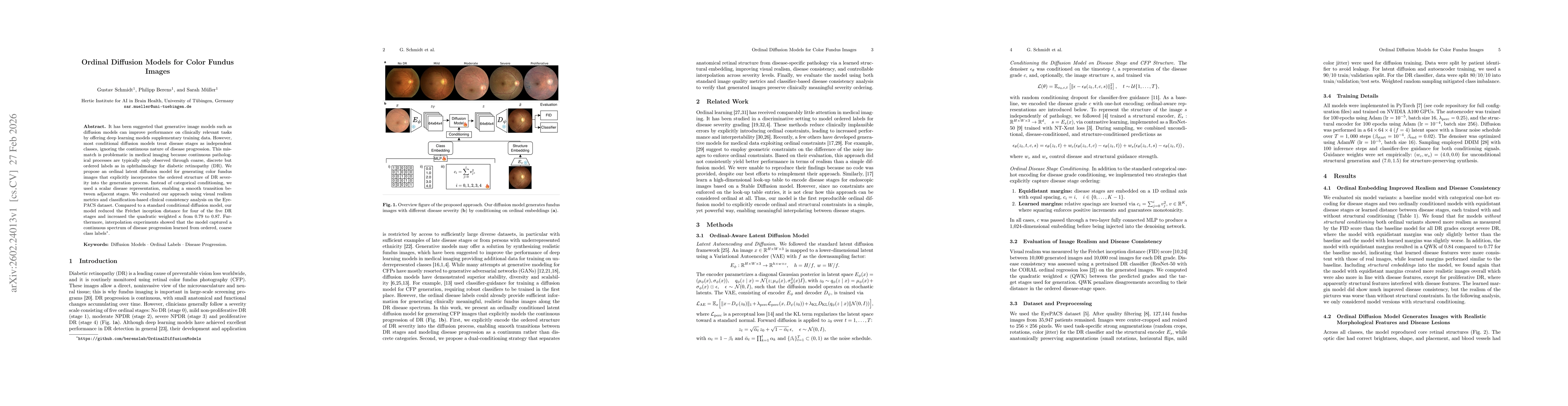
It has been suggested that generative image models such as diffusion models can improve performance on clinically relevant tasks by offering deep learning models supplementary training data. However, most conditional diffusion models treat disease stages as independent classes, ignoring the continuous nature of disease progression. This mismatch is problematic in medical imaging because continuous pathological processes are typically only observed through coarse, discrete but ordered labels as in ophthalmology for diabetic retinopathy (DR). We propose an ordinal latent diffusion model for generating color fundus images that explicitly incorporates the ordered structure of DR severity into the generation process. Instead of categorical conditioning, we used a scalar disease representation, enabling a smooth transition between adjacent stages. We evaluated our approach using visual realism metrics and classification-based clinical consistency analysis on the EyePACS dataset. Compared to a standard conditional diffusion model, our model reduced the Fréchet inception distance for four of the five DR stages and increased the quadratic weighted $κ$ from 0.79 to 0.87. Furthermore, interpolation experiments showed that the model captured a continuous spectrum of disease progression learned from ordered, coarse class labels.
AI Key Findings
Generated Mar 02, 2026
Methodology — What approach did the authors take?
Ordinal latent diffusion model conditioned on a scalar disease representation using equidistant or learned ordinal embeddings, combined with a structural encoder for anatomical conditioning; trained on the EyePACS fundus dataset and evaluated with FID and quadratic weighted kappa (QWK) metrics
Key Results — What are the main findings?
- Ordinal conditioning reduced FID for four of five diabetic retinopathy stages and raised QWK from 0.79 to 0.87, indicating improved realism and disease consistency
- Equidistant ordinal margins yielded the best overall performance, while learned margins improved disease consistency but slightly degraded realism
- Interpolation across the ordinal axis produced smooth transitions of pathological features, demonstrating the model’s ability to capture a continuous disease spectrum
Significance — Why does this research matter?
Provides a method to generate realistic, disease‑stage‑aware fundus images that can augment training data, enable counterfactual analyses, and improve understanding of disease progression in ophthalmology
Technical Contribution — What is the technical contribution?
Development of an ordinal latent diffusion framework that embeds disease stages on a continuous axis, incorporates structural conditioning, and demonstrates continuous progression modeling via interpolation
Novelty — What is new about this work?
First work to explicitly encode ordinal disease stages in diffusion models for medical imaging, combining ordinal embeddings with structural conditioning and counterfactual generation
Limitations — What are the limitations of this study?
- Reliance on automated metrics (FID and classifier‑based QWK) limits assessment of true clinical correctness
- Artifacts and reduced realism in advanced stages (proliferative DR) indicate challenges in modeling complex pathology
Future Work — What did the authors propose for future work?
- Integrate expert clinical evaluation and higher‑resolution synthesis to capture fine pathological details
- Extend the framework to longitudinal datasets to model temporal disease progression
How to Cite This Paper
@article{schmidt2026ordinal,
title = {Ordinal Diffusion Models for Color Fundus Images},
author = {Schmidt, Gustav and Berens, Philipp and Müller, Sarah},
year = {2026},
eprint = {2602.24013},
archivePrefix = {arXiv},
primaryClass = {cs.CV},
}
Schmidt, G., Berens, P., & Müller, S. (2026). Ordinal Diffusion Models for Color Fundus Images. arXiv. https://arxiv.org/abs/2602.24013
Schmidt, Gustav, et al. "Ordinal Diffusion Models for Color Fundus Images." arXiv, 2026, arxiv.org/abs/2602.24013.
PDF Preview
Similar Papers
Found 4 papersVascX Models: Deep Ensembles for Retinal Vascular Analysis From Color Fundus Images.
van Garderen, Karin A, Vargas Quiros, Jose, Liefers, Bart et al.
VascX Models: Model Ensembles for Retinal Vascular Analysis from Color Fundus Images
Eyened Reading Center, Sinergia Consortium, Jose Vargas Quiros et al.
Generation of Structurally Realistic Retinal Fundus Images with Diffusion Models
Soochahn Lee, Sojung Go, Younghoon Ji et al.
Generating Realistic Counterfactuals for Retinal Fundus and OCT Images using Diffusion Models
Indu Ilanchezian, Valentyn Boreiko, Laura Kühlewein et al.
Comments (0)