This paper proposes a novel orthogonal annotation method for semi-supervised 3D medical image segmentation, which labels only two orthogonal slices to reduce annotation effort. It employs a Dense-Sparse Co-training (DeSCO) model to leverage both dense and sparse pseudo labels, achieving high segmentation accuracy with minimal annotations.
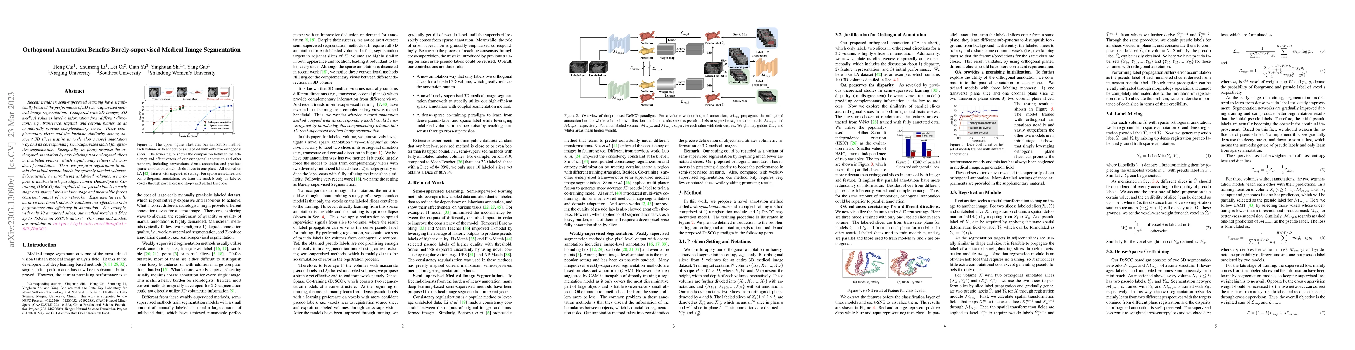
Recent trends in semi-supervised learning have significantly boosted the performance of 3D semi-supervised medical image segmentation. Compared with 2D images, 3D medical volumes involve information from different directions, e.g., transverse, sagittal, and coronal planes, so as to naturally provide complementary views. These complementary views and the intrinsic similarity among adjacent 3D slices inspire us to develop a novel annotation way and its corresponding semi-supervised model for effective segmentation. Specifically, we firstly propose the orthogonal annotation by only labeling two orthogonal slices in a labeled volume, which significantly relieves the burden of annotation. Then, we perform registration to obtain the initial pseudo labels for sparsely labeled volumes. Subsequently, by introducing unlabeled volumes, we propose a dual-network paradigm named Dense-Sparse Co-training (DeSCO) that exploits dense pseudo labels in early stage and sparse labels in later stage and meanwhile forces consistent output of two networks. Experimental results on three benchmark datasets validated our effectiveness in performance and efficiency in annotation. For example, with only 10 annotated slices, our method reaches a Dice up to 86.93% on KiTS19 dataset.
Get AI-generated insights about this paper's methodology, results, and significance.
Current paper (gray), citations (green), references (blue)
Zhiqiang Shen, Peng Cao, Junming Su et al.
Xiaomeng Li, Kwang-Ting Cheng, Yiqun Lin et al.
Banafshe Felfeliyan, Jacob L. Jaremko, Abhilash Hareendranathan et al.
Zhang Yi, Wang Yan, Wang Lituan et al.
Title | Authors | Year | Actions |
---|---|---|---|
Barely-Supervised Brain Tumor Segmentation via Employing Segment Anything Model
|
Yuhang Ding, Hongmin Liu
|
2025
|
View Paper |
Focus-TransUnet3D: High-Precision Model for 3D Segmentation of Medical Point Targets
|
Dihua Zhai, Hao Li, Qingyuan Liu, Ke Tian, Yi Yang, Zhenyao Chang, Shuo Wang, Yuanqing Xia
|
2025
|
View Paper |
Leveraging Textual Anatomical Knowledge for Class-Imbalanced Semi-Supervised Multi-Organ Segmentation
|
Yuliang Gu, Weilun Tsao, Bo Du, Thierry G'eraud, Yongchao Xu
|
2025
|
View Paper |
PMT: Progressive Mean Teacher via Exploring Temporal Consistency for Semi-Supervised Medical Image Segmentation
|
Ning Gao, Sanping Zhou, Le Wang, Nanning Zheng
|
2024
|
View Paper |
On the Viability of Semi-Supervised Segmentation Methods for Statistical Shape Modeling
|
Janmesh Ukey, Tushar Kataria, Shireen Elhabian
|
2024
|
View Paper |
The Devil is in the Statistics: Mitigating and Exploiting Statistics Difference for Generalizable Semi-supervised Medical Image Segmentation
|
Muyang Qiu, Jian Zhang, Lei Qi, Qian Yu, Yinghuan Shi, Yang Gao
|
2024
|
View Paper |
Self-Paced Sample Selection for Barely-Supervised Medical Image Segmentation
|
Junming Su, Zhiqiang Shen, Peng Cao, Jinzhu Yang, Osmar R. Zaïane
|
2024
|
View Paper |
Bi-Fusion of Structure and Deformation at Multi-Scale for Joint Segmentation and Registration
|
Jiaju Zhang, Tianyu Fu, Deqiang Xiao, Jingfan Fan, Hong Song, Danni Ai, Jian Yang
|
2024
|
View Paper |
Rethinking Barely-Supervised Volumetric Medical Image Segmentation from an Unsupervised Domain Adaptation Perspective
|
Zhiqiang Shen, Peng Cao, Junming Su, Jinzhu Yang, Osmar R. Zaiane
|
2024
|
View Paper |
ToNNO: Tomographic Reconstruction of a Neural Network's Output for Weakly Supervised Segmentation of 3D Medical Images
|
Marius Schmidt-Mengin, Alexis Benichoux, S. Belachew, N. Komodakis, Nikos Paragios
|
2024
|
View Paper |
Constructing and Exploring Intermediate Domains in Mixed Domain Semi-supervised Medical Image Segmentation
|
Qinghe Ma, Jian Zhang, Lei Qi, Qian Yu, Yinghuan Shi, Yang Gao
|
2024
|
View Paper |
Estimation and Analysis of Slice Propagation Uncertainty in 3D Anatomy Segmentation
|
Rachaell Nihalaani, Tushar Kataria, Jadie Adams, Shireen Elhabian
|
2024
|
View Paper |
Semi-supervised Medical Image Segmentation through Dual-subnet Mutual Correction
|
Mei Sun, Jintao Li, Lulu He, Xuemei Sun
|
2024
|
View Paper |
3D Medical Image Segmentation with Sparse Annotation via Cross-Teaching between 3D and 2D Networks
|
Heng Cai, Lei Qi, Qian Yu, Yinghuan Shi, Yang Gao
|
2023
|
View Paper |
SAMMed: A medical image annotation framework based on large vision model
|
Chenglong Wang, Dexuan Li, Sucheng Wang, Chengxiu Zhang, Yida Wang, Yun Liu, Guang Yang
|
2023
|
View Paper |
3-D Seismic Fault Detection Using Fault Orthogonal Annotation and Barely Supervised Learning
|
Zheng Zhang, Zhe Yan, Haiying Li, Jiankun Jing, Hao Yan, Hanming Gu
|
2025
|
View Paper |
FM-ABS: Promptable Foundation Model Drives Active Barely Supervised Learning for 3D Medical Image Segmentation
|
Zhe Xu, Cheng Chen, Donghuan Lu, Jinghan Sun, Dong Wei, Yefeng Zheng, Quanzheng Li, R. Tong
|
2024
|
View Paper |
3-D Salt Body Segmentation Method Based on Multiview Co-Regularization
|
Zhifeng Xu, Kewen Li, Zongchao Huang, Ruonan Yin, Yating Fan
|
2024
|
View Paper |
Decoupled Training for Semi-supervised Medical Image Segmentation with Worst-Case-Aware Learning
|
Ankit Das, Chandan Gautam, Hisham Cholakkal, Pritee Agrawal, Feng Yang, Ramasamy Savitha, Yong Liu
|
2024
|
View Paper |
Gradient-Aware for Class-Imbalanced Semi-supervised Medical Image Segmentation
|
Wenbo Qi, Jiafei Wu, S. Chan
|
2024
|
View Paper |
Comments (0)