This paper addresses the challenges of detecting cancer in whole slide images (WSIs) by overcoming limitations of patch-based learning. It proposes a binary cancer detection network for post neoadjuvant therapy breast cancer WSIs, emphasizing the need for sensitivity and precision across the entire slide, and introduces a negative data sampling strategy that significantly reduces false positives and improves detection accuracy.
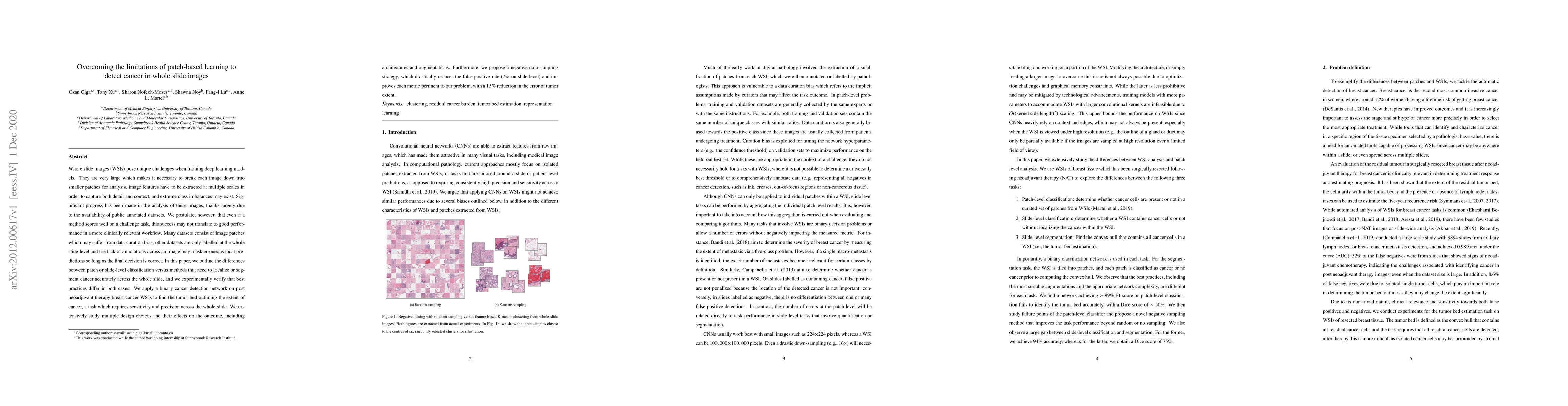
Whole slide images (WSIs) pose unique challenges when training deep learning models. They are very large which makes it necessary to break each image down into smaller patches for analysis, image features have to be extracted at multiple scales in order to capture both detail and context, and extreme class imbalances may exist. Significant progress has been made in the analysis of these images, thanks largely due to the availability of public annotated datasets. We postulate, however, that even if a method scores well on a challenge task, this success may not translate to good performance in a more clinically relevant workflow. Many datasets consist of image patches which may suffer from data curation bias; other datasets are only labelled at the whole slide level and the lack of annotations across an image may mask erroneous local predictions so long as the final decision is correct. In this paper, we outline the differences between patch or slide-level classification versus methods that need to localize or segment cancer accurately across the whole slide, and we experimentally verify that best practices differ in both cases. We apply a binary cancer detection network on post neoadjuvant therapy breast cancer WSIs to find the tumor bed outlining the extent of cancer, a task which requires sensitivity and precision across the whole slide. We extensively study multiple design choices and their effects on the outcome, including architectures and augmentations. Furthermore, we propose a negative data sampling strategy, which drastically reduces the false positive rate (7% on slide level) and improves each metric pertinent to our problem, with a 15% reduction in the error of tumor extent.
Get AI-generated insights about this paper's methodology, results, and significance.
Current paper (gray), citations (green), references (blue)
Emiel A. M. Janssen, Kjersti Engan, Neel Kanwal et al.
Title | Authors | Year | Actions |
---|---|---|---|
Segmentation of cortical bone, trabecular bone, and medullary pores from micro-CT images using 2D and 3D deep learning models.
|
Andrew H Lee, Julian M Moore, Brandon Vera Covarrubias, Leigha M. Lynch
|
2025
|
View Paper |
Diagnostic classification in toxicologic pathology using attention-guided weak supervision and whole slide image features: a pilot study in rat livers.
|
Philip Zehnder, Jeffrey Feng, Trung Nguyen, Philip Shen, R. Sullivan, Reina N Fuji, Fangyao Hu
|
2025
|
View Paper |
Vision Transformers for Low-Quality Histopathological Images: A Case Study on Squamous Cell Carcinoma Margin Classification
|
So-yun Park, Gelan Ayana, Beshatu Debela Wako, Kwangcheol Casey Jeong, Soon‐Do Yoon, Se-woon Choe
|
2025
|
View Paper |
A universal immunohistochemistry analyzer for generalizing AI-driven assessment of immunohistochemistry across immunostains and cancer types
|
B. Brattoli, Mohammad Mostafavi, Taebum Lee, W. Jung, J. Ryu, Seonwook Park, Jongchan Park, Sergio Pereira, Seunghwan Shin, Sangjoon Choi, Hyojin Kim, D. Yoo, S. Ali, K. Paeng, C. Ock, Soo Ick Cho, Seokhwi Kim
|
2024
|
View Paper |
Non-Generative Artificial Intelligence (AI) in Medicine: Advancements and Applications in Supervised and Unsupervised Machine Learning.
|
Liron Pantanowitz, Thomas Pearce, Ibrahim Abukhiran, Matthew G. Hanna, Sarah Wheeler, T. R. Soong, Ahmad P. Tafti, Joshua Pantanowitz, Ming Y. Lu, Faisal Mahmood, Qiangqiang Gu, Hooman H. Rashidi
|
2024
|
View Paper |
Modified U-Net based Framework for Laparoscopic Procesures
|
Kumar Mayank, Anchal Kumar, Pushp Jain, Vipin Pal, Yogita
|
2024
|
View Paper |
Çekişmeli Üretici Ağlar Kullanılarak Medikal Görüntülerin Sanal Hematoksilen ve Eozin (H&E) Boyanması
|
M. Aydın
|
2024
|
View Paper |
Distilling High Diagnostic Value Patches for Whole Slide Image Classification Using Attention Mechanism
|
Tianhang Nan, Hao Quan, Yong Ding, Xingyu Li, Kai Yang, Xiaoyu Cui
|
2024
|
View Paper |
Artificial Intelligence Models for the Detection of Microsatellite Instability from Whole-Slide Imaging of Colorectal Cancer
|
Gavino Faa, Ferdinando Coghe, A. Pretta, Massimo Castagnola, Peter Van Eyken, Luca Saba, M. Scartozzi, Matteo Fraschini
|
2024
|
View Paper |
URCDM: Ultra-Resolution Image Synthesis in Histopathology
|
Sarah Cechnicka, James Ball, Matthew Baugh, Hadrien Reynaud, Naomi Simmonds, A. Smith, Catherine Horsfield, C. Roufosse, Bernhard Kainz
|
2024
|
View Paper |
Deep Residual Learning-Based Classification with Identification of Incorrect Predictions and Quantification of Cellularity and Nuclear Morphological Features in Digital Pathological Images of Common Astrocytic Tumors
|
Yen-Chang Chen, Shinn-Zong Lin, Jia-Ru Wu, Wei-Hsiang Yu, H. Harn, Wen-Chiuan Tsai, Ching-Ann Liu, Ken-Leiang Kuo, Chao-Yuan Yeh, Sheng-Tzung Tsai
|
2024
|
View Paper |
STimage-1K4M: A histopathology image-gene expression dataset for spatial transcriptomics
|
Jiawen Chen, Muqing Zhou, Wenrong Wu, Jinwei Zhang, Yun Li, Didong Li
|
2024
|
View Paper |
Model-Agnostic Binary Patch Grouping for Bone Marrow WSI Representation.
|
Youqing Mu, H. R. Tizhoosh, Taher Dehkharghanian, Saghir Alfasly, Clinton J. V. Campbell
|
2024
|
View Paper |
Prediction of Tuberculosis From Lung Tissue Images of Diversity Outbred Mice Using Jump Knowledge Based Cell Graph Neural Network
|
Vasundhara Acharya, Diana Choi, B. Yener, G. Beamer
|
2024
|
View Paper |
Seeing the random forest through the decision trees. Supporting learning health systems from histopathology with machine learning models: Challenges and opportunities
|
Ricardo Gonzalez, Ashirbani Saha, Clinton J.V. Campbell, Peyman Nejat, Cynthia Lokker, Andrew P. Norgan
|
2023
|
View Paper |
Gravity Network for end-to-end small lesion detection
|
Ciro Russo, Alessandro Bria, Claudio Marrocco
|
2023
|
View Paper |
Deep Architecture for Breast Cancer Detection: Using Optimized VGG16 Model and Transfer Learning
|
Akash Chauhan, Indrajeet Kumar, K. Singh, Prabhdeep Singh
|
2023
|
View Paper |
DHUnet: Dual-branch hierarchical global-local fusion network for whole slide image segmentation
|
Lian-min Wang, Liangrui Pan, Hetian Wang, Mingting Liu, Zhichao Feng, Pengfei Rong, Zuo Chen, Shaoliang Peng
|
2023
|
View Paper |
Whole Slide Images in Artificial Intelligence Applications in Digital Pathology: Challenges and Pitfalls
|
K. Başak, K. Ozyoruk, D. Demir
|
2023
|
View Paper |
Probabilistic Attention Based on Gaussian Processes for Deep Multiple Instance Learning
|
Arne Schmidt, Pablo Morales-Álvarez, R. Molina
|
2023
|
View Paper |
An AI-based approach for detecting cells and microbial byproducts in low volume scanning electron microscope images of biofilms
|
Dilanga L. B. Abeyrathna, M. Ashaduzzaman, M. Malshe, Jawahar Kalimuthu, V. Gadhamshetty, P. Chundi, M. Subramaniam
|
2022
|
View Paper |
Simulation Palynologists for Pollinosis Prevention: A Progressive Learning of Pollen Localization and Classification for Whole Slide Images
|
Linna Zhao, Jianqiang Li, Wenhui Cheng, Su-Qin Liu, Zhengkai Gao, Xi Xu, Caihua Ye, Huanling You
|
2022
|
View Paper |
Computer-Aided Cancer Diagnosis via Machine Learning and Deep Learning: A comparative review
|
Solene Bechelli
|
2022
|
View Paper |
Bench to bedside: recent advances in lung cancer pathological research with implications for diagnostic clinical practice
|
D. Moore, J. Le Quesne
|
2022
|
View Paper |
Identify Representative Samples by Conditional Random Field of Cancer Histology Images
|
Yiqing Shen, Xiaoyao Liang, D. Shen, Jing Ke
|
2022
|
View Paper |
Influence of uncertainty estimation techniques on false-positive reduction in liver lesion detection
|
Ishaan Bhat, J. Pluim, M. Viergever, H. Kuijf
|
2022
|
View Paper |
Fusing pre-trained convolutional neural networks features for multi-differentiated subtypes of liver cancer on histopathological images
|
Xiaogang Dong, Min Li, Panyun Zhou, Xin Deng, Siyue Li, Xingyue Zhao, Yi Wu, J. Qin, Wenjia Guo
|
2022
|
View Paper |
Deep contrastive learning based tissue clustering for annotation-free histopathology image analysis
|
Jiangpeng Yan, Hanbo Chen, Xiu Li, Jianhua Yao
|
2022
|
View Paper |
Collaborative Federated Learning behind Hospitals' Firewalls for Predicting Histological Response to Neoadjuvant Chemotherapy in Triple-Negative Breast Cancer
|
J. Ogier du Terrail, A. Leopold, C. Joly, C. Béguier, M. Andreux, C. Maussion, B. Schmauch, Eric W. Tramel, E. Bendjebbar, M. Zaslavskiy, G. Wainrib, M. Milder, J. Gervasoni, J. Guérin, T. Durand, A. Livartowski, K. Moutet, C. Gautier, I. Djafar, A.-L. Moisson, C. Marini, M. Galtier, G. Bataillon, P. Heudel
|
2021
|
View Paper |
Improving Self-supervised Learning with Hardness-aware Dynamic Curriculum Learning: An Application to Digital Pathology
|
C. Srinidhi, Anne L. Martel
|
2021
|
View Paper |
Self supervised contrastive learning for digital histopathology
|
Ozan Ciga, Anne L. Martel, Tony Xu
|
2020
|
View Paper |
Classification of breast pathological images based on multiscale information interaction and fusion
|
Weilong Ding, Fenglong Zhu, Zheng Kui, Xiupeng Jia
|
2024
|
View Paper |
Volume-based Performance not Guaranteed by Promising Patch-based Results in Medical Imaging
|
Abhishek Moturu, Sayali Joshi, A. Doria, A. Goldenberg
|
2022
|
View Paper |
Hyper-AdaC: Adaptive clustering-based hypergraph representation of whole slide images for survival analysis
|
Hakim Benkirane, M. Vakalopoulou, S. Christodoulidis, I. Garberis, S. Michiels, P. Cournède
|
2022
|
View Paper |
Comments (0)