Papanicolaou Stain Unmixing for RGB Image Using Weighted Nucleus Sparsity and Total Variation Regularization
Publication
Metrics
AI Quick Summary
This research presents a novel method for quantifying dyes in Papanicolaou-stained RGB images, overcoming previous challenges with multispectral imaging. The approach, validated against multispectral results, uses optimization with nonnegativity, sparsity, and total variation regularizations. It successfully distinguished precancerous lesions, achieving 98.0% accuracy, showcasing RGB-based stain unmixing's potential for quantitative cervical cancer screening.
Paper Preview
Abstract
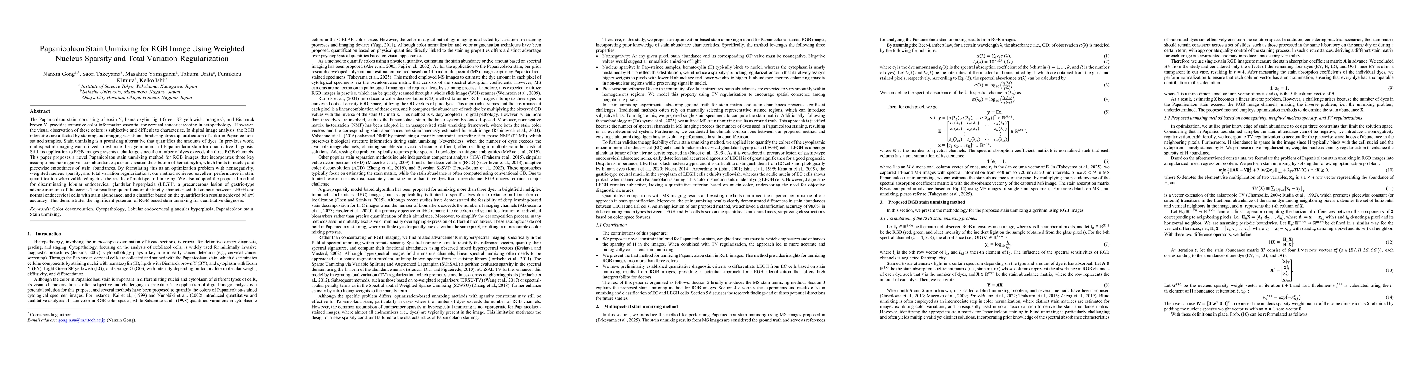
The Papanicolaou stain, consisting of eosin Y, hematoxylin, light Green SF yellowish, orange G, and Bismarck brown Y, provides extensive color information essential for cervical cancer screening in cytopathology. However, the visual observation of these colors is subjective and difficult to characterize. In digital image analysis, the RGB intensities are affected by staining and imaging variations, hindering direct quantification of color in Papanicolaou-stained samples. Stain unmixing is a promising alternative that quantifies the amounts of dyes. In previous work, multispectral imaging was utilized to estimate the dye amounts of Papanicolaou stain for quantitative diagnosis. Still, its application to RGB images presents a challenge since the number of dyes exceeds the three RGB channels. This paper proposes a novel Papanicolaou stain unmixing method for RGB images that incorporates three key assumptions: nonnegative stain abundances; a sparse spatial distribution of hematoxylin, which binds to nuclei; and piecewise smoothness of stain abundances. By formulating this as an optimization problem with nonnegativity, weighted nucleus sparsity, and total variation regularizations, our method achieved excellent performance in stain quantification when validated against the results of multispectral imaging. We also adopted the proposed method for discriminating lobular endocervical glandular hyperplasia (LEGH), a precancerous lesion of gastric-type adenocarcinoma of the cervix. The resulting quantification distinctly characterized differences between LEGH and normal endocervical cells with stain abundance, and a classifier based on the quantification results achieved 98.0% accuracy. This demonstrates the significant potential of RGB-based stain unmixing for quantitative diagnosis.
AI Key Findings
Get AI-generated insights about this paper's methodology, results, significance, and more — seven facets brought into focus.
Impact
Paper Details
Authors
PDF Preview
Citation Network
Current paper (gray), citations (green), references (blue)
Display is limited for performance on very large graphs.
Discussion 0