Authors
Summary
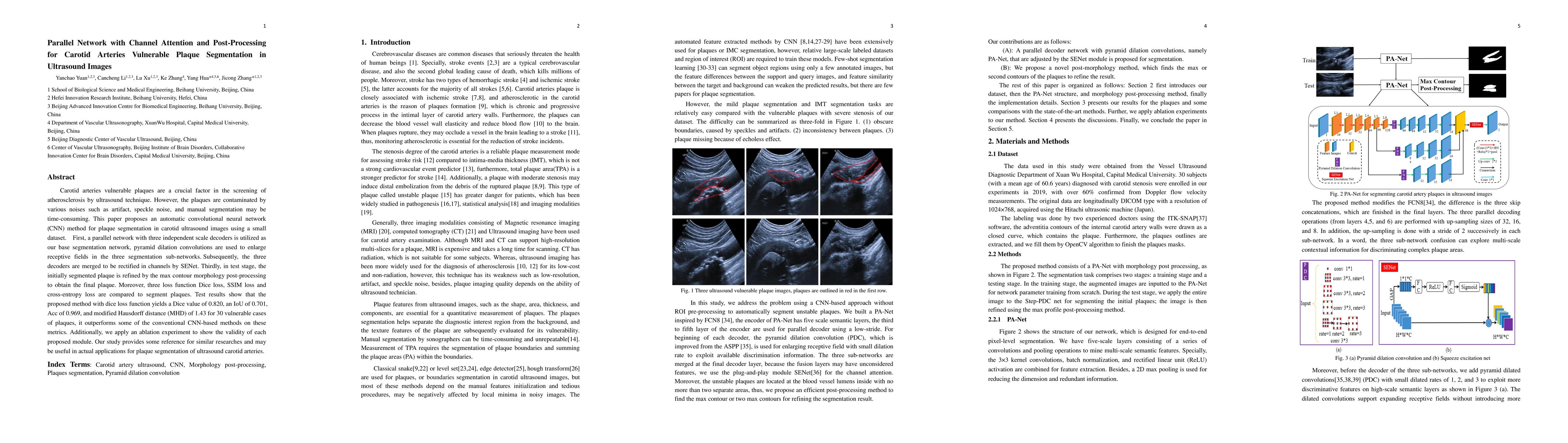
Carotid arteries vulnerable plaques are a crucial factor in the screening of atherosclerosis by ultrasound technique. However, the plaques are contaminated by various noises such as artifact, speckle noise, and manual segmentation may be time-consuming. This paper proposes an automatic convolutional neural network (CNN) method for plaque segmentation in carotid ultrasound images using a small dataset. First, a parallel network with three independent scale decoders is utilized as our base segmentation network, pyramid dilation convolutions are used to enlarge receptive fields in the three segmentation sub-networks. Subsequently, the three decoders are merged to be rectified in channels by SENet. Thirdly, in test stage, the initially segmented plaque is refined by the max contour morphology post-processing to obtain the final plaque. Moreover, three loss function Dice loss, SSIM loss and cross-entropy loss are compared to segment plaques. Test results show that the proposed method with dice loss function yields a Dice value of 0.820, an IoU of 0.701, Acc of 0.969, and modified Hausdorff distance (MHD) of 1.43 for 30 vulnerable cases of plaques, it outperforms some of the conventional CNN-based methods on these metrics. Additionally, we apply an ablation experiment to show the validity of each proposed module. Our study provides some reference for similar researches and may be useful in actual applications for plaque segmentation of ultrasound carotid arteries.
AI Key Findings
Get AI-generated insights about this paper's methodology, results, and significance.
Paper Details
PDF Preview
Key Terms
Citation Network
Current paper (gray), citations (green), references (blue)
Display is limited for performance on very large graphs.
Similar Papers
Found 4 papersCarotid Plaque Segmentation in Ultrasound Images Using a Mask R-CNN
Tomy Varghese, Maxwell J. Kiernan, Rashid Al Mukaddim et al.
DualPlaqueNet with dual-branch structure and attention mechanism for carotid plaque semantic segmentation and size prediction.
Deng, Lili, Duan, Xiaokai, Wang, Yunling et al.
Automatic Multi-Task Segmentation and Vulnerability Assessment of Carotid Plaque on Contrast-Enhanced Ultrasound Images and Videos via Deep Learning.
Zhang, Han, Chen, Jing, He, Da et al.
| Title | Authors | Year | Actions |
|---|
Comments (0)