Publication
Metrics
AI Quick Summary
This paper proposes transfer learning methods for analyzing myopic fundus images, achieving top ranks in the Pathologic Myopia Challenge for tasks like classification, fovea localization, and optic disc segmentation. The success is attributed to adapting established deep learning architectures.
Paper Preview
Abstract
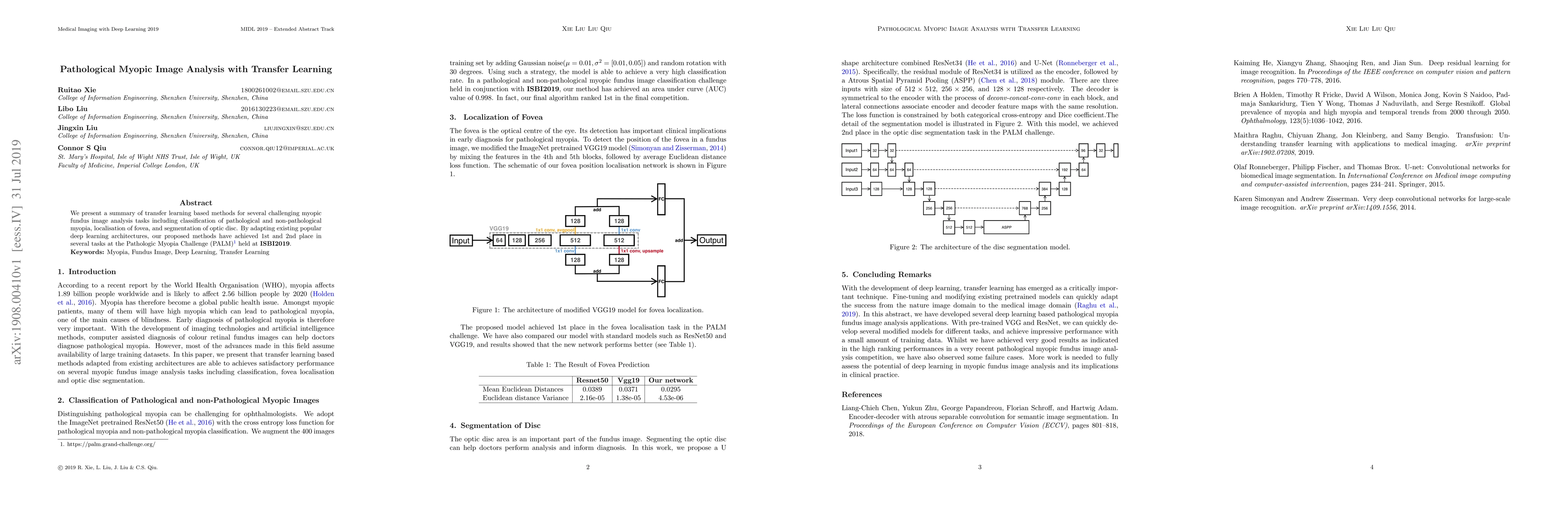
We present a summary of transfer learning based methods for several challenging myopic fundus image analysis tasks including classification of pathological and non-pathological myopia,localisation of fovea,and segmentation of optic disc.By adapting existing popular deep learning architectures,our proposed methods have achieved 1st and 2nd place in several tasks at the Pathologic Myopia Challenge held at ISBI2019.
AI Key Findings
Get AI-generated insights about this paper's methodology, results, significance, and more — seven facets brought into focus.
Impact
Paper Details
PDF Preview
Key Terms
Citation Network
Current paper (gray), citations (green), references (blue)
Display is limited for performance on very large graphs.
Discussion 0