Pathological OCT Retinal Layer Segmentation using Branch Residual U-shape Networks
Publication
Metrics
AI Quick Summary
This paper introduces a novel CNN architecture combining dilated residual blocks in an asymmetric U-shape network for segmenting retinal layers in OCT imaging, especially for late-stage eye diseases. The proposed method shows lower computational costs and higher performance compared to existing techniques, validated on a dataset of late-stage AMD patients.
Paper Preview
Abstract
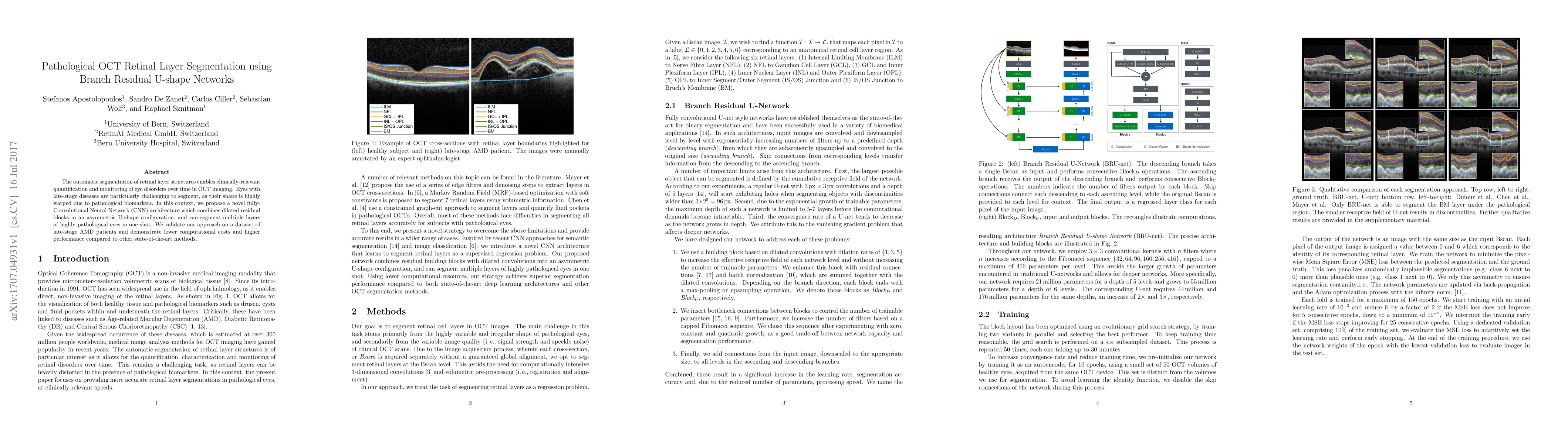
The automatic segmentation of retinal layer structures enables clinically-relevant quantification and monitoring of eye disorders over time in OCT imaging. Eyes with late-stage diseases are particularly challenging to segment, as their shape is highly warped due to pathological biomarkers. In this context, we propose a novel fully Convolutional Neural Network (CNN) architecture which combines dilated residual blocks in an asymmetric U-shape configuration, and can segment multiple layers of highly pathological eyes in one shot. We validate our approach on a dataset of late-stage AMD patients and demonstrate lower computational costs and higher performance compared to other state-of-the-art methods.
AI Key Findings
Get AI-generated insights about this paper's methodology, results, significance, and more — seven facets brought into focus.
Impact
Paper Details
PDF Preview
Key Terms
Citation Network
Current paper (gray), citations (green), references (blue)
Display is limited for performance on very large graphs.
Discussion 0