Pathology-Guided AI System for Accurate Segmentation and Diagnosis of Cervical Spondylosis

Source: ArXiv

AI Quick Summary

This paper presents an AI-assisted Expert-based Diagnosis System for automated segmentation and diagnosis of cervical spondylosis using MRI, achieving an average Dice coefficient of over 0.90 and a mean absolute error of 2.44 degrees for Cobb angle measurement, outperforming existing methods.

Share: Share on X LinkedIn

Summary

Cervical spondylosis, a complex and prevalent condition, demands precise and efficient diagnostic techniques for accurate assessment. While MRI offers detailed visualization of cervical spine anatomy, manual interpretation remains labor-intensive and prone to error. To address this, we developed an innovative AI-assisted Expert-based Diagnosis System that automates both segmentation and diagnosis of cervical spondylosis using MRI. Leveraging a dataset of 960 cervical MRI images from patients with cervical disc herniation, our system 
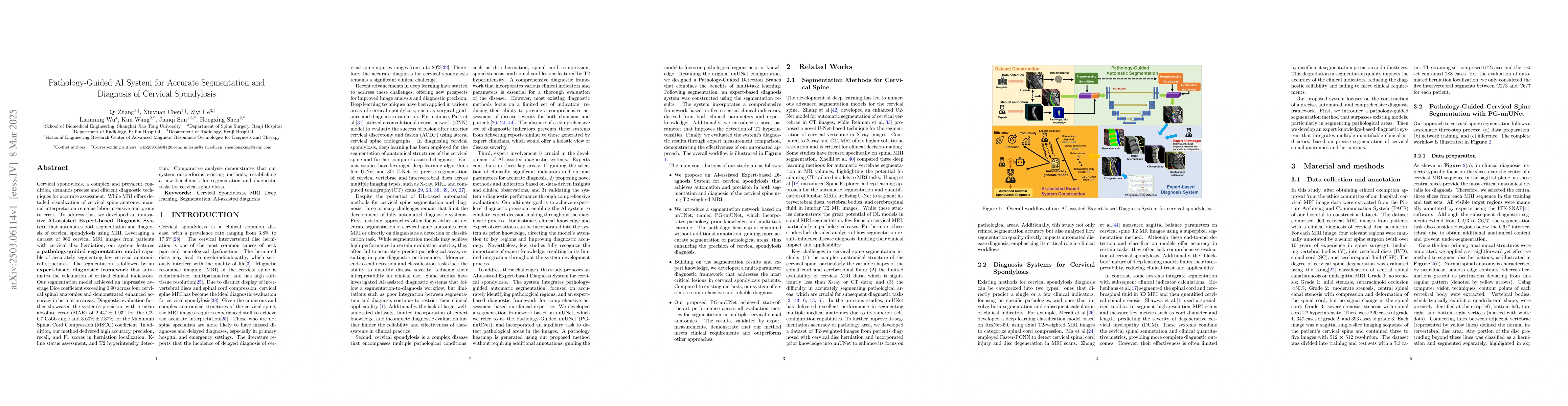
features a pathology-guided segmentation model capable of accurately segmenting key cervical anatomical structures. The segmentation is followed by an expert-based diagnostic framework that automates the calculation of critical clinical indicators. Our segmentation model achieved an impressive average Dice coefficient exceeding 0.90 across four cervical spinal anatomies and demonstrated enhanced accuracy in herniation areas. Diagnostic evaluation further showcased the system precision, with a mean absolute error (MAE) of 2.44 degree for the C2-C7 Cobb angle and 3.60 precentage for the Maximum Spinal Cord Compression (MSCC) coefficient. In addition, our method delivered high accuracy, precision, recall, and F1 scores in herniation localization, K-line status assessment, and T2 hyperintensity detection. Comparative analysis demonstrates that our system outperforms existing methods, establishing a new benchmark for segmentation and diagnostic tasks for cervical spondylosis.

AI Key Findings

Generated Jun 10, 2025

Methodology

The research developed an AI-assisted Expert-based Diagnosis System for cervical spondylosis using MRI, featuring a pathology-guided segmentation model and an expert-based diagnostic framework.

Key Results

  • Segmentation model achieved an average Dice coefficient exceeding 0.90 across four cervical spinal anatomies.
  • Mean absolute error (MAE) of 2.44 degrees for the C2-C7 Cobb angle and 3.60 percentage for the Maximum Spinal Cord Compression (MSCC) coefficient.
  • High accuracy, precision, recall, and F1 scores in herniation localization, K-line status assessment, and T2 hyperintensity detection.
  • Outperformed existing methods, establishing a new benchmark for segmentation and diagnostic tasks for cervical spondylosis.

Significance

This research provides a reliable, automated solution for cervical spondylosis diagnosis, enhancing efficiency and accuracy compared to manual interpretation, which is labor-intensive and prone to error.

Technical Contribution

The Pathology-Guided AI System leverages a novel PG-nnUNet architecture, integrating edge loss and a Pathology-Guided Detection Branch for enhanced segmentation accuracy, particularly in pathological regions.

Novelty

The proposed system outperforms traditional CNN-based and Transformer-based models in both segmentation and diagnostic tasks, offering a reliable, automated solution for cervical spondylosis diagnosis with expert-level performance.

Limitations

  • The study utilized only T2-weighted MRI images; future work will incorporate multi-parametric MRI, X-ray, and CT data for a more comprehensive multi-modality diagnostic system.
  • The dataset was sourced from a single hospital, and future research should integrate external datasets from various hospitals and research centers for broader generalizability.

Future Work

  • Expand the dataset to include multi-modal imaging data for improved diagnostic performance.
  • Incorporate axial MRI images for comprehensive segmentation and clinical indicator calculation.

Paper Details

PDF Preview

Download PDF

Citation Network

Current paper (gray), citations (green), references (blue)

Display is limited for performance on very large graphs.

Current Paper
Citations
References
Click to view

No citations found for this paper.

Comments (0)

This website uses cookies to ensure you get the best experience. Learn more