Quick Summary:

This paper introduces PCMC-T1, a physically-constrained deep-learning model for free-breathing myocardial T1 mapping, addressing the challenge of respiratory motion artifacts. PCMC-T1 outperforms baseline methods in model fitting quality and clinical impact, while maintaining comparable anatomical alignment.

Summary

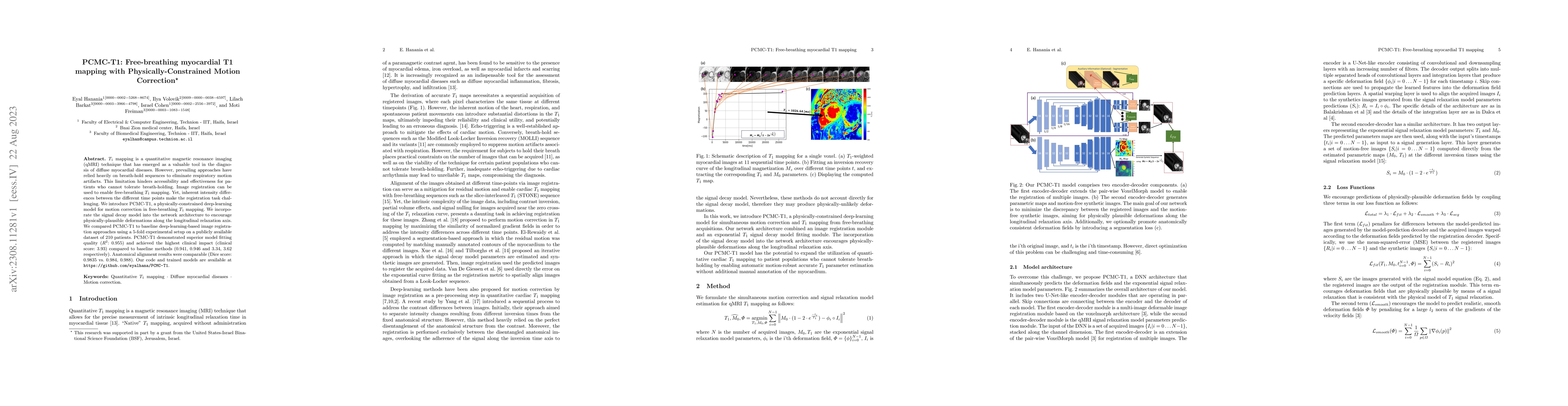
T1 mapping is a quantitative magnetic resonance imaging (qMRI) technique that has emerged as a valuable tool in the diagnosis of diffuse myocardial diseases. However, prevailing approaches have relied heavily on breath-hold sequences to eliminate respiratory motion artifacts. This limitation hinders accessibility and effectiveness for patients who cannot tolerate breath-holding. Image registration can be used to enable free-breathing T1 mapping. Yet, inherent intensity differences between the different time points make the registration task challenging. We introduce PCMC-T1, a physically-constrained deep-learning model for motion correction in free-breathing T1 mapping. We incorporate the signal decay model into the network architecture to encourage physically-plausible deformations along the longitudinal relaxation axis. We compared PCMC-T1 to baseline deep-learning-based image registration approaches using a 5-fold experimental setup on a publicly available dataset of 210 patients. PCMC-T1 demonstrated superior model fitting quality (R2: 0.955) and achieved the highest clinical impact (clinical score: 3.93) compared to baseline methods (0.941, 0.946 and 3.34, 3.62 respectively). Anatomical alignment results were comparable (Dice score: 0.9835 vs. 0.984, 0.988). Our code and trained models are available at https://github.com/eyalhana/PCMC-T1.

AI Key Findings

Get AI-generated insights about this paper's methodology, results, and significance.

Paper Details

PDF Preview

Download PDF

Key Terms

mapping (0.197)
patients (0.189)
clinical (0.188)
physically (0.182)
score (0.180)
baseline (0.165)
deep learning (0.154)
motion (0.134)
image (0.130)
diseases (0.126)
deep (0.126)
network architecture (0.123)
available (0.123)
learning model (0.122)
approaches (0.121)
artifacts (0.120)
plausible (0.120)
vs (0.120)
free (0.119)
diagnosis (0.118)

Citation Network

Current paper (gray), citations (green), references (blue)

Current Paper
Citations
References
Click to view
Title Authors Year Actions
Contrast-insensitive motion correction for MRI cardiac T1 mapping
Chengyu Yue, Lu Huang, Lihong Huang, Yi Guo, Qian Tao, Liming Xia, Yuanyuan Wang
2025
View Paper
Enhanced DTCMR With Cascaded Alignment and Adaptive Diffusion
Fanwen Wang, Yihao Luo, Camila Munoz, Ke Wen, Yaqing Luo, Jiahao Huang, Yinzhe Wu, Z. Khalique, Maria Molto, R. Rajakulasingam, Ranil de Silva, D. Pennell, Pedro F. Ferreira, Andrew D. Scott, S. Nielles-Vallespin, Guang Yang
2024
View Paper
MBSS-T1: Model-based subject-specific self-supervised motion correction for robust cardiac T1 mapping
Eyal Hanania, Adi Zehavi-Lenz, Ilya Volovik, Daphna Link-Sourani, Israel Cohen, Moti Freiman
2024
View Paper
CardioSpectrum: Comprehensive Myocardium Motion Analysis with 3D Deep Learning and Geometric Insights
Shahar Zuler, Shai Tejman-Yarden, Dan Raviv
2024
View Paper
Groupwise Deformable Registration of Diffusion Tensor Cardiovascular Magnetic Resonance: Disentangling Diffusion Contrast, Respiratory and Cardiac Motions
Fanwen Wang, Yihao Luo, Ke Wen, Jiahao Huang, Pedro F. Ferreira, Yaqing Luo, Yinzhe Wu, Camila Munoz, D. Pennell, Andrew D. Scott, S. Nielles-Vallespin, Guang Yang
2024
View Paper
Deep-learning-based groupwise registration for motion correction of cardiac T1 mapping
Yi Zhang, Yidong Zhao, Lu Huang, Li Xia, Qian Tao
2024
View Paper

Comments (0)

This website uses cookies to ensure you get the best experience. Learn more