Publication
Metrics
AI Quick Summary
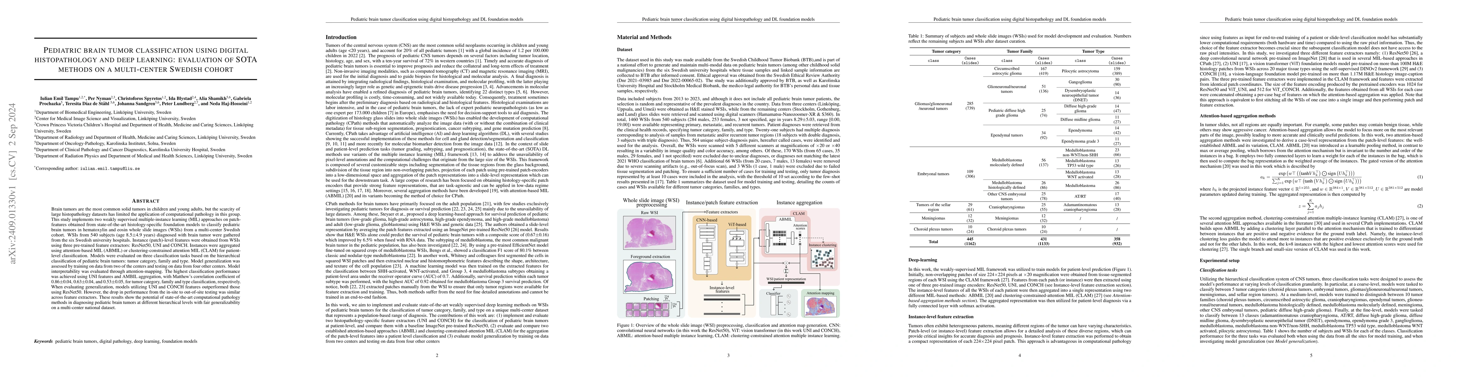
This study employs deep learning and multiple-instance learning to classify pediatric brain tumors using histopathology images from a large multi-center Swedish cohort. The best performing model, using UNI features and ABMIL aggregation, demonstrated high classification accuracy and fair generalizability across different hospitals.
Paper Preview
Abstract
Brain tumors are the most common solid tumors in children and young adults, but the scarcity of large histopathology datasets has limited the application of computational pathology in this group. This study implements two weakly supervised multiple-instance learning (MIL) approaches on patch-features obtained from state-of-the-art histology-specific foundation models to classify pediatric brain tumors in hematoxylin and eosin whole slide images (WSIs) from a multi-center Swedish cohort. WSIs from 540 subjects (age 8.5$\pm$4.9 years) diagnosed with brain tumor were gathered from the six Swedish university hospitals. Instance (patch)-level features were obtained from WSIs using three pre-trained feature extractors: ResNet50, UNI and CONCH. Instances were aggregated using attention-based MIL (ABMIL) or clustering-constrained attention MIL (CLAM) for patient-level classification. Models were evaluated on three classification tasks based on the hierarchical classification of pediatric brain tumors: tumor category, family and type. Model generalization was assessed by training on data from two of the centers and testing on data from four other centers. Model interpretability was evaluated through attention-mapping. The highest classification performance was achieved using UNI features and AMBIL aggregation, with Matthew's correlation coefficient of 0.86$\pm$0.04, 0.63$\pm$0.04, and 0.53$\pm$0.05, for tumor category, family and type classification, respectively. When evaluating generalization, models utilizing UNI and CONCH features outperformed those using ResNet50. However, the drop in performance from the in-site to out-of-site testing was similar across feature extractors. These results show the potential of state-of-the-art computational pathology methods in diagnosing pediatric brain tumors at different hierarchical levels with fair generalizability on a multi-center national dataset.
AI Key Findings
Get AI-generated insights about this paper's methodology, results, significance, and more — seven facets brought into focus.
Impact
Authors
PDF Preview
Citation Network
Current paper (gray), citations (green), references (blue)
Display is limited for performance on very large graphs.
Discussion 0