01
MethodologyHow they did it
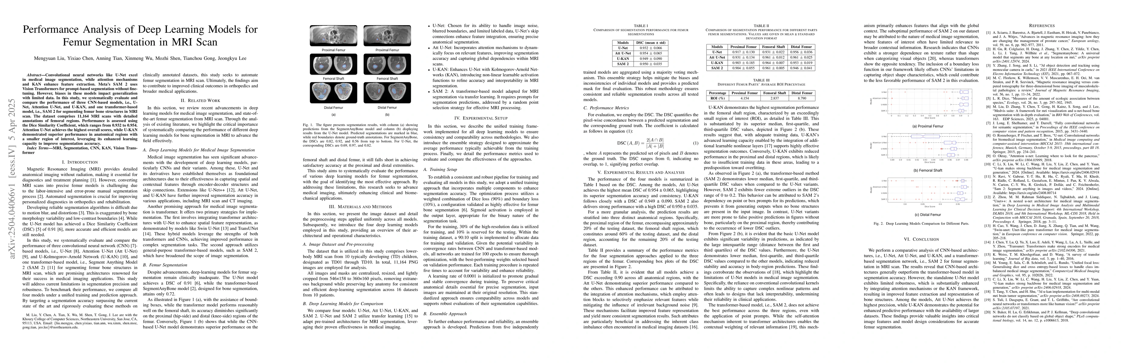
This study systematically evaluates and compares the performance of three CNN-based models (U-Net, Attention U-Net, U-KAN) and one transformer-based model (SAM 2) for femur segmentation in MRI scans using a dataset of 11,164 scans with detailed annotations. Performance is assessed using the Dice Similarity Coefficient (DSC) ranging from 0.932 to 0.954.
Discussion 0