Perfusion Imaging and Single Material Reconstruction in Polychromatic Photon Counting CT
Publication
Metrics
Paper Preview
Abstract
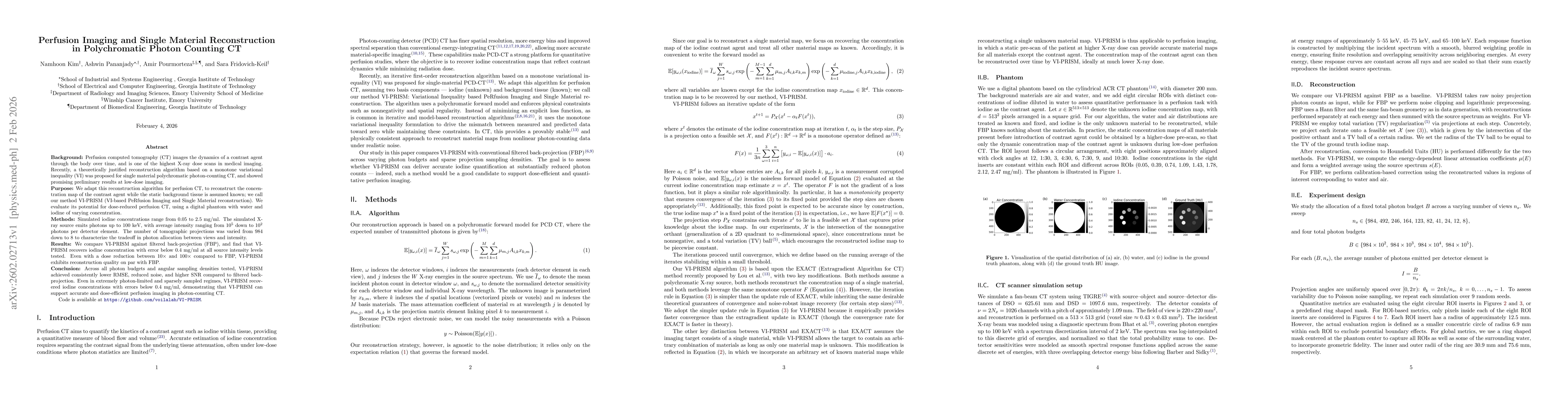
Background: Perfusion computed tomography (CT) images the dynamics of a contrast agent through the body over time, and is one of the highest X-ray dose scans in medical imaging. Recently, a theoretically justified reconstruction algorithm based on a monotone variational inequality (VI) was proposed for single material polychromatic photon-counting CT, and showed promising early results at low-dose imaging. Purpose: We adapt this reconstruction algorithm for perfusion CT, to reconstruct the concentration map of the contrast agent while the static background tissue is assumed known; we call our method VI-PRISM (VI-based PeRfusion Imaging and Single Material reconstruction). We evaluate its potential for dose-reduced perfusion CT, using a digital phantom with water and iodine of varying concentration. Methods: Simulated iodine concentrations range from 0.05 to 2.5 mg/ml. The simulated X-ray source emits photons up to 100 keV, with average intensity ranging from $10^5$ down to $10^2$ photons per detector element. The number of tomographic projections was varied from 984 down to 8 to characterize the tradeoff in photon allocation between views and intensity. Results: We compare VI-PRISM against filtered back-projection (FBP), and find that VI-PRISM recovers iodine concentration with error below 0.4 mg/ml at all source intensity levels tested. Even with a dose reduction between 10x and 100x compared to FBP, VI-PRISM exhibits reconstruction quality on par with FBP. Conclusion: Across all photon budgets and angular sampling densities tested, VI-PRISM achieved consistently lower RMSE, reduced noise, and higher SNR compared to filtered back-projection. Even in extremely photon-limited and sparsely sampled regimes, VI-PRISM recovered iodine concentrations with errors below 0.4 mg/ml, showing that VI-PRISM can support accurate and dose-efficient perfusion imaging in photon-counting CT.
AI Key Findings
Get AI-generated insights about this paper's methodology, results, significance, and more — seven facets brought into focus.
Discussion 0