Summary
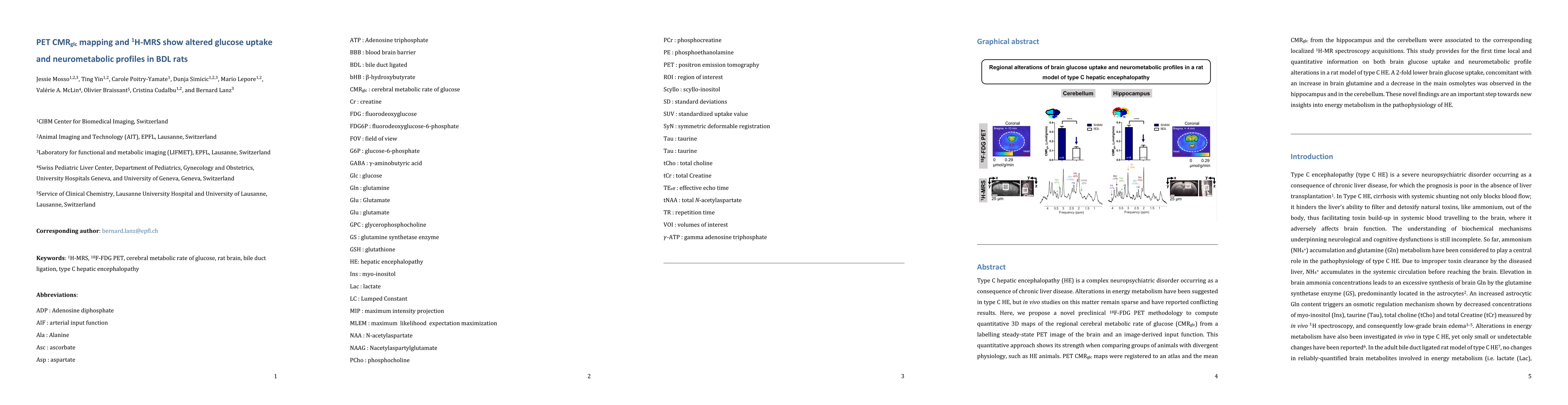
Type C hepatic encephalopathy (HE) is a complex neuropsychiatric disorder occurring as a consequence of chronic liver disease. Alterations in energy metabolism have been suggested in type C HE, but $\textit{in vivo}$ studies on this matter remain sparse and have reported conflicting results. Here, we propose a novel preclinical $^{18}$F-FDG PET methodology to compute quantitative 3D maps of the regional cerebral metabolic rate of glucose (CMR$_{glc}$) from a labelling steady-state PET image of the brain and an image-derived input function. This quantitative approach shows its strength when comparing groups of animals with divergent physiology, such as HE animals. PET CMR$_{glc}$ maps were registered to an atlas and the mean CMR$_{glc}$ from the hippocampus and the cerebellum were associated to the corresponding localized $^{1}$H MR spectroscopy acquisitions. This study provides for the first time local and quantitative information on both brain glucose uptake and neurometabolic profile alterations in a rat model of type C HE. A 2-fold lower brain glucose uptake, concomitant with an increase in brain glutamine and a decrease in the main osmolytes was observed in the hippocampus and in the cerebellum. These novel findings are an important step towards new insights into energy metabolism in the pathophysiology of HE.
AI Key Findings
Get AI-generated insights about this paper's methodology, results, and significance.
Paper Details
PDF Preview
Key Terms
Citation Network
Current paper (gray), citations (green), references (blue)
Display is limited for performance on very large graphs.
Similar Papers
Found 4 papersMapping of neurovascular and neurometabolic couplings by multimodal optical imaging.
Sanganahalli, Basavaraju G, Herman, Peter, Sanggaard, Simon et al.
Associations between Aβ deposition and neurometabolic alterations in Alzheimer's disease: Insights from hybrid 3D MRSI-PET imaging.
Liu, Jun, Zhang, Miao, Li, Yao et al.
Impact of hyperglycemia and antidiabetic medication on pancreatic uptake on [68Ga]Ga-DOTATOC PET/CT.
Kunte, Sophie Carina, Siegmund, Thorsten, Tiling, Maximilian et al.
| Title | Authors | Year | Actions |
|---|
Comments (0)