This paper introduces oblique back-illumination microscopy (OBM) to extend phase contrast techniques like differential interference contrast (DIC) to thick biological samples, enabling near video-rate in vivo phase imaging suitable for endoscopic applications.
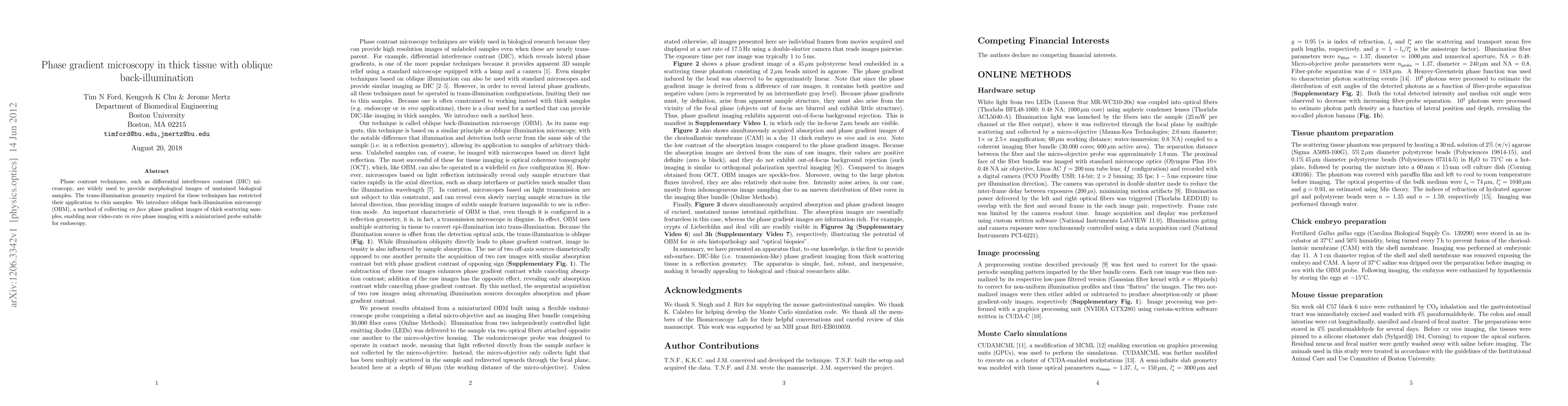
Phase contrast techniques, such as differential interference contrast (DIC) microscopy, are widely used to provide morphological images of unstained biological samples. The trans-illumination geometry required for these techniques has restricted their application to thin samples. We introduce oblique back-illumination microscopy (OBM), a method of collecting en face phase gradient images of thick scattering samples, enabling near video-rate in vivo phase imaging with a miniaturized probe suitable for endoscopy.
This graph shows how this paper is connected to other research. The current paper is shown in dark gray, papers that cite it are in green, and papers it references are in blue. You can:
• Hover over any node to see the paper title
• Click a node to view that paper's details
• Drag nodes to rearrange the graph
• Use the zoom controls (bottom right) to zoom in/out
Title | Authors | Year | Actions |
---|---|---|---|
Label-free quantitative imaging of conjunctival goblet cells
|
Noseong Park, Suil Jeon, Seonghan Kim, Jungbin Lee, Jin Suk Ryu, Wan Jae Choi, Chang Ho Yoon, Chulmin Joo, Ki Hean Kim
|
2025
|
View Paper |
Quantitative phase imaging endoscopy with a metalens
|
A. Shanker, Johannes E. Fröch, Saswata Mukherjee, Maksym V. Zhelyeznyakov, Steve Brunton, Eric J Seibel, Arka Majumdar
|
2024
|
View Paper |
Ultrasensitive in vivo infrared spectroscopic imaging via oblique photothermal microscopy
|
Mingsheng Li, Sheng Xiao, Hongli Ni, Guangrui Ding, Yuhao Yuan, Carolyn Marar, Jerome Mertz, Ji-Xin Cheng
|
2024
|
View Paper |
Novel imaging and biophysical approaches to study tissue hydraulics in mammalian folliculogenesis
|
Jake Turley, Kim Whye Leong, C. J. Chan
|
2024
|
View Paper |
Non-invasive label-free imaging analysis pipeline for in situ characterization of 3D brain organoids
|
Caroline E Serafini, Seleipiri Charles, Paloma Casteleiro Costa, Weibo Niu, Brian F. Cheng, Zhexing Wen, Hang Lu, Francisco E. Robles
|
2024
|
View Paper |
New Avenues for Phase Imaging: Optical Metasurfaces
|
N. Priscilla, S. Sulejman, Ann Roberts, L. Wesemann
|
2024
|
View Paper |
Quantitative phase imaging based on holography: trends and new perspectives
|
Zhengzhong Huang, Liangcai Cao
|
2024
|
View Paper |
Quantitative phase imaging by gradient retardance optical microscopy.
|
Jinming Zhang, M. Sarollahi, Shirley Luckhart, Maria J. Harrison, Andreas E. Vasdekis
|
2024
|
View Paper |
Non-Invasive Label-free Analysis Pipeline for In Situ Characterization of Differentiation in 3D Brain Organoid Models
|
Caroline Filan, Seleipiri Charles, Paloma Casteleiro Costa, Weibo Niu, Brian F. Cheng, Zhexing Wen, Hang Lu, Francisco E. Robles
|
2024
|
View Paper |
Perspective on quantitative phase imaging to improve precision cancer medicine
|
Yang Liu, S. Uttam
|
2024
|
View Paper |
High-contrast and high-speed multimodal imaging platform: the adaptive optics-confocal rolling slit ophthalmoscope (AO-CRSO)
|
L. Krafft, P. Senée, O. Thouvenin, D. Castro Farías, M. Paques, P. Mecê, S. Meimon
|
2024
|
View Paper |
Tracking cellular and subcellular dynamics with quantitative oblique back-illumination microscopy
|
Caroline Filan, Srinidhi Bharadwaj, Paloma Casteleiro Costa, Amin Davarzani, D. Cappabianca, Anna Tommasi, Lauren Sarko, Nina La Vonne Denne, Leidong Mao, Krishanu Saha, L. Karumbaiah, Francisco E. Robles
|
2024
|
View Paper |
Label-free functional analysis of root-associated microbes with dynamic quantitative oblique back-illumination microscopy
|
Caroline E Serafini, Madison Green, Abigail Diering, M. Cicerone, Lily S. Cheung, J. Kostka, Francisco E. Robles
|
2024
|
View Paper |
Improved resolution in fiber bundle inline holographic microscopy using multiple illumination sources
|
Michael R. Hughes, Callum McCall
|
2024
|
View Paper |
High-speed, phase contrast retinal and blood flow imaging using an adaptive optics partially confocal multi-line ophthalmoscope
|
Soohyun Lee, Stacey S. Choi, R. Meleppat, Robert J. Zawadzki, Nathan Doble
|
2024
|
View Paper |
Motion Contrast, Phase Gradient, and Simultaneous OCT Images Assist in the Interpretation of Dark-Field Images in Eyes with Retinal Pathology
|
M. Mujat, Konstantina Sampani, Ankit H. Patel, Ronald Zambrano, Jennifer K Sun, G. Wollstein, R. D. Ferguson, J. Schuman, N. Iftimia
|
2024
|
View Paper |
Novel aspects of signal processing in lamina I
|
B. V. Safronov, P. Szucs
|
2024
|
View Paper |
Cellular-Level Analysis of Retinal Blood Vessel Walls Based on Phase Gradient Images
|
M. Mujat, Konstantina Sampani, Ankit H. Patel, Jennifer K Sun, N. Iftimia
|
2023
|
View Paper |
Quantitative Phase Imaging with a Metalens
|
A. Shanker, Johannes E. Froech, Saswata Mukherjee, Maksym V. Zhelyeznyakov, Eric J Seibel, Arka Majumdar
|
2023
|
View Paper |
Analysis of structural effects of sickle cell disease on brain vasculature of mice using three-dimensional quantitative phase imaging
|
Caroline Filan, Hannah Song, Manu O Platt, Francisco E. Robles
|
2023
|
View Paper |
In vivo optical trapping of erythrocytes in mouse liver imaged with oblique back-illumination microscopy
|
Meng Shao, Rui Liu, Changxu Li, Zhenhao Chai, Zhensheng Zhong, Fengya Lu, Xunbin Wei, Jinhua Zhou, Min-Cheng Zhong
|
2023
|
View Paper |
Miniaturized microscope for non-invasive imaging of leukocyte-endothelial interaction in human microcirculation
|
A. Bagramyan, Charles P. Lin
|
2023
|
View Paper |
The Rotational Doppler Effect of Twisted Photons in Scattered Fields
|
Yanwang Zhai, Jingtao Fan, Hui Qiao, Tiankuang Zhou, Jiamin Wu, Qionghai Dai
|
2023
|
View Paper |
Quantitative phase imaging of opaque specimens with flexible endoscopic microscopy
|
Jingyi Wang, Wu You, Yuheng Jiao, Yanhong Zhu, Xiaojun Liu, X. Jiang, Chenfei Hu, Wenlong Lu
|
2023
|
View Paper |
Imaging and qualitative depth analysis with a portable nonmydriatic fundus camera using oblique illumination
|
N. Kaushik, Takashi Sasaki, Kana Takeyama, K. Hane, T. Nakazawa
|
2023
|
View Paper |
Mode conversion of qOBM (quantitative oblique back-illumination microscopy) stain-free tissue images to emulate H&E histology via deep learning
|
T. Abraham, P. C. Costa, Caroline Filan, Francisco E. Robles, R. Levenson
|
2023
|
View Paper |
Single capture quantitative phase imaging with tomographic sectioning
|
Paloma Casteleiro Costa, Z. Guang, Nischita Kaza, Francisco E. Robles
|
2023
|
View Paper |
Quantitative phase imaging of sickle cell disease effects on mouse brain vasculature using quantitative oblique back-illumination microscopy
|
Caroline Filan, Hannah W. Song, M. Platt, Francisco E. Robles
|
2023
|
View Paper |
Tracking early tuberous sclerosis complex diseased organoid development with quantitative oblique back-illumination microscopy
|
Caroline Filan, Seleipiri Charles, Paloma Casteleiro Costa, B. Cheng, Hang Lu, Francisco E. Robles
|
2023
|
View Paper |
Asymmetrical Illumination Enables Lipid Droplets Segmentation in Caenorhabditis elegans Using Epi-Illumination Dark Field Microscopy
|
Ruijie Shi, Yu Sun, J. Fang, Xiangyang Chen, Zachary J Smith, K. Chu
|
2022
|
View Paper |
Functional imaging with dynamic quantitative oblique back-illumination microscopy
|
Paloma Casteleiro Costa, Bryan Wang, Caroline Filan, Annie Bowles-Welch, C. Yeago, Krishnendu Roy, Francisco E. Robles
|
2022
|
View Paper |
Label-free multiscale dynamic imaging using 3D phase contrast and deep UV microscopy
|
Francisco E. Robles
|
2022
|
View Paper |
Roadmap on wavefront shaping and deep imaging in complex media
|
S. Gigan, O. Katz, H. D. Aguiar, E. Andresen, A. Aubry, J. Bertolotti, E. Bossy, D. Bouchet, Joshua Brake, S. Brasselet, Y. Bromberg, Hui Cao, Thomas Chaigne, Zhongtao Cheng, W. Choi, Tom'avs vCivzm'ar, Meng Cui, Vincent R. Curtis, H. Defienne, M. Hofer, R. Horisaki, R. Horstmeyer, Na Ji, Aaron K. LaViolette, J. Mertz, C. Moser, A. Mosk, Nicolas C. P'egard, R. Piestun, S. Popoff, D. Phillips, D. Psaltis, Babak Rahmani, H. Rigneault, S. Rotter, L. Tian, I. Vellekoop, L. Waller, Lihong Wang, Timothy D. Weber, Sheng Xiao, Chris Xu, A. Yamilov, Changhuei Yang, H. Yılmaz
|
2021
|
View Paper |
Design of a radial multi-offset detection pattern for in vivo phase contrast imaging of the inner retina in humans.
|
Elena Gofas-Salas, Yuhua Rui, P. Mecê, M. Zhang, V. Snyder, K. Vienola, Daniel M W Lee, J. Sahel, K. Grieve, E. Rossi
|
2021
|
View Paper |
Epi‐illumination dark‐field microscopy enables direct visualization of unlabeled small organisms with high spatial and temporal resolution
|
Ruijie Shi, Xiangyang Chen, Jing Huo, Siyue Guo, Zachary J Smith, K. Chu
|
2021
|
View Paper |
Single shot quantitative phase gradient estimation using Wigner-Ville distribution in digital holographic microscopy
|
Ankur Vishnoi, R. Gannavarpu
|
2021
|
View Paper |
High-speed multifocus phase imaging in thick tissue
|
Sheng Xiao, Shuqi Zheng, J. Mertz
|
2021
|
View Paper |
Epi-illumination quantitative phase imaging (QPI) and 3D refractive index (RI) tomography in thick scattering samples with quantitative oblique back-illumination microscopy
|
Francisco E. Robles
|
2021
|
View Paper |
Epi-illumination tomographic quantitative phase imaging identifies brain tumor margins
|
Paloma Casteleiro Costa, Z. Guang, P. Ledwig, Zhaobin Zhang, S. Neill, J. Olson, Francisco E. Robles
|
2021
|
View Paper |
Quantitative phase imaging of thick scattering samples through a flexible fiber-optic bundle
|
Z. Guang, P. Ledwig, Paloma Casteleiro Costa, Francisco E. Robles
|
2021
|
View Paper |
Towards in-vivo label-free detection of brain tumor margins with epi-illumination tomographic quantitative phase imaging.
|
P. C. Costa, Z. Guang, P. Ledwig, Zhaobin Zhang, S. Neill, J. Olson, Francisco E. Robles
|
2021
|
View Paper |
Real-time holographic lensless micro-endoscopy through flexible fibers via fiber bundle distal holography
|
Noam Badt, O. Katz
|
2021
|
View Paper |
Design of a radial multi-offset detection pattern for in vivo phase contrast imaging of the retinal ganglion cells in humans
|
Elena Gofas-Salas, Yuhua Rui, P. Mecê, Min Zhang, V. Snyder, K. Vienola, Daniel M W Lee, J. Sahel, Kate Grieve, E. Rossi
|
2020
|
View Paper |
Quantitative 3D refractive index tomography of opaque samples in epi-mode.
|
P. Ledwig, Francisco E. Robles
|
2020
|
View Paper |
Anti-aliasing high resolution quantitative phase microscopy based on differential phase contrast imaging
|
Yao Fan, Jiasong Sun, Qian Chen, Chao Zuo
|
2020
|
View Paper |
Label-free microendoscopy using a micro-needle imaging probe for in vivo deep tissue imaging.
|
Kwanjun Park, June Hoan Kim, Taedong Kong, Woong Sun, Jonghwan Lee, T. Yang, Youngwoon Choi
|
2020
|
View Paper |
Inline holographic microscopy through fiber imaging bundles.
|
M. Hughes
|
2020
|
View Paper |
LED array reflectance microscopy for scattering-based multi-contrast imaging.
|
Weiye Song, Alex Matlock, Sipei Fu, Xiaodan Qin, Hui Feng, C. Gabel, L. Tian, Ji Yi
|
2020
|
View Paper |
In vivo corneal and lenticular microscopy with asymmetric fundus retroillumination
|
Timothy D. Weber, J. Mertz
|
2020
|
View Paper |
Epi-mode tomographic quantitative phase imaging in thick scattering samples
|
Francisco E. Robles
|
2020
|
View Paper |
Spheroscope: A custom-made miniaturized microscope for tracking tumour spheroids in microfluidic devices
|
A. Rodríguez-Pena, J. Uranga-Solchaga, C. Ortíz-de-Solórzano, Iván Cortés-Domínguez
|
2020
|
View Paper |
Optimizing white blood cell contrast in graded-field capillaroscopy using capillary tissue phantoms
|
Gregory N. McKay, Taylor L. Bobrow, Srivathsan Kalyan, S. Hur, N. Durr
|
2020
|
View Paper |
3D refractive index tomography with quantitative oblique back-illumination microscopy
|
P. Ledwig, Francisco E. Robles
|
2020
|
View Paper |
Origin of cell contrast in offset aperture adaptive optics ophthalmoscopy.
|
Andres Guevara-Torres, David R. Williams, Jesse Schallek
|
2020
|
View Paper |
Noninvasive white blood cell quantification in umbilical cord blood collection bags with quantitative oblique back‐illumination microscopy
|
Paloma Casteleiro Costa, P. Ledwig, Austin Bergquist, J. Kurtzberg, Francisco E. Robles
|
2020
|
View Paper |
Imaging human blood cells in vivo with oblique back-illumination capillaroscopy.
|
Gregory N. McKay, N. Mohan, N. Durr
|
2020
|
View Paper |
Inverse scattering for reflection intensity phase microscopy.
|
Alex Matlock, A. Sentenac, P. Chaumet, Ji Yi, L. Tian
|
2019
|
View Paper |
Epi-illumination gradient light interference microscopy for imaging opaque structures
|
M. Kandel, Chenfei Hu, G. Naseri Kouzehgarani, Eunjung Min, Kathryn M. Sullivan, H. Kong, Jennifer M. Li, D. Robson, M. Gillette, C. Best-Popescu, G. Popescu
|
2019
|
View Paper |
Epi-mode tomographic quantitative phase imaging in thick scattering samples.
|
P. Ledwig, Francisco E. Robles
|
2019
|
View Paper |
Transscleral Optical Phase Imaging of the Human Retina
|
T. Laforest, Mathieu Künzi, L. Kowalczuk, D. Carpentras, F. Behar-Cohen, C. Moser
|
2019
|
View Paper |
Label-free 3D imaging of weakly absorbing samples using spatially-incoherent annular illumination microscopy.
|
Xiao Ma, Bowen Zhou, Zijian Su, Zibang Zhang, Junzheng Peng, Jingang Zhong
|
2019
|
View Paper |
Bond-selective transient phase imaging via sensing of the infrared photothermal effect
|
Delong Zhang, L. Lan, Yeran Bai, Hassaan Majeed, M. Kandel, G. Popescu, Ji‐Xin Cheng
|
2018
|
View Paper |
Quantitative phase and polarization imaging through an optical fiber applied to detection of early esophageal tumorigenesis
|
G. Gordon, J. Joseph, M. Alcolea, T. Sawyer, Calum Williams, C. R. M. Fitzpatrick, P. Jones, M. di Pietro, R. Fitzgerald, T. Wilkinson, S. Bohndiek
|
2018
|
View Paper |
Quantitative phase and polarisation endoscopy applied to detection of early oesophageal tumourigenesis
|
G. Gordon, J. Joseph, M. Alcolea, T. Sawyer, Alexander J. Macfaden, Calum Williams, C. R. M. Fitzpatrick, P. Jones, M. Pietro, R. Fitzgerald, T. Wilkinson, S. Bohndiek
|
2018
|
View Paper |
Wide-field anti-aliased quantitative differential phase contrast microscopy.
|
Yao Fan, Jiasong Sun, Qian Chen, Jianqin Zhang, Chao Zuo
|
2018
|
View Paper |
Pupil plane differential detection microscopy.
|
H. Paudel, C. Alt, J. Runnels, Charles P. Lin
|
2018
|
View Paper |
Image computing for fibre-bundle endomicroscopy: A review
|
A. Perperidis, K. Dhaliwal, S. Mclaughlin, Tom Kamiel Magda Vercauteren
|
2018
|
View Paper |
Dual-wavelength oblique back-illumination microscopy for the non-invasive imaging and quantification of blood in collection and storage bags.
|
P. Ledwig, Moses Sghayyer, J. Kurtzberg, Francisco E. Robles
|
2018
|
View Paper |
Non-mydriatic chorioretinal imaging in a transmission geometry and application to retinal oximetry
|
Timothy D. Weber, J. Mertz
|
2018
|
View Paper |
Retina and Choroid Imaging with Transcranial Back-illumination
|
Timothy D. Weber, J. Mertz
|
2018
|
View Paper |
In vivo flow cytometry for blood cell analysis using differential epi-detection of forward scattered light
|
H. Paudel, Yookyung Jung, A. Raphael, C. Alt, Juwell W. Wu, Judith Runnels, Charles P. Lin
|
2018
|
View Paper |
A dual-mode mobile phone microscope using the onboard camera flash and ambient light
|
A. Orth, Emma R Wilson, Jeremy G. Thompson, B. Gibson
|
2018
|
View Paper |
A new microscopy for imaging retinal cells
|
T. Laforest, D. Carpentras, Mathieu Kunzi, L. Kowalczuk, F. Behar-Cohen, C. Moser
|
2017
|
View Paper |
Optofluidic time-stretch quantitative phase microscopy.
|
Baoshan Guo, Cheng Lei, Denny Wu, H. Kobayashi, Takuro Ito, Yaxiaer Yalikun, Sangwook Lee, A. Isozaki, Ming Li, Yiyue Jiang, A. Yasumoto, D. Di Carlo, Yo Tanaka, Y. Yatomi, Y. Ozeki, K. Goda
|
2017
|
View Paper |
Smartphone-based multi-contrast microscope using color-multiplexed illumination
|
Daeseong Jung, Jun-Ho Choi, Soocheol Kim, Suho Ryu, Wonchan Lee, Jong-Seok Lee, C. Joo
|
2017
|
View Paper |
A dual-mode mobile phone microscope using integrated and ambient light
|
A. Orth, Emma R Wilson, Jeremy G. Thompson, B. Gibson
|
2017
|
View Paper |
Optical 3D imaging of subcellular dynamics in biological cultures and tissues : applications to ophthalmology and neuroscience
|
O. Thouvenin
|
2017
|
View Paper |
DMD based digital speckle illumination for high resolution imaging
|
Anant B. Shinde, A. Mishra, S. M. Perinchery, V. M. Murukeshan
|
2017
|
View Paper |
Holotomography: refractive index as an intrinsic imaging contrast for 3-D label-free live cell imaging
|
Doyeon Kim, Sangyun Lee, Moosung Lee, Juntaek Oh, Su-A Yang, Yongkeun Park
|
2017
|
View Paper |
Quantitative phase imaging of retinal cells
|
T. Laforest, D. Carpentras, L. Kowalczuk, F. Behar-Cohen, C. Moser
|
2017
|
View Paper |
Widefield fluorescence microscopy with sensor-based conjugate adaptive optics using oblique back illumination
|
Jiang Li, T. Bifano, J. Mertz
|
2016
|
View Paper |
Dual-modality endomicroscopy with co-registered fluorescence and phase contrast.
|
Cong Ba, M. Palmiere, Jason T. Ritt, Jerome Mertz
|
2016
|
View Paper |
Vascular Fragility and the Endothelial Glycocalyx in the Tissues Lining the Healthy Gingival Crevice.
|
D. Townsend, F. D’Aiuto, J. Deanfield
|
2016
|
View Paper |
Tyndall Windows: Tunable Scattering of Disordered Solid–Liquid Matching Mixtures
|
Benedikt Groever, B. Heshmat, R. Raskar
|
2016
|
View Paper |
3D differential phase contrast microscopy
|
Michael Chen, L. Tian, L. Waller
|
2016
|
View Paper |
Invited Review Article: Pump-probe microscopy.
|
M. Fischer, Jesse W. Wilson, Francisco E. Robles, W. Warren
|
2016
|
View Paper |
Optical brush: Imaging through permuted probes
|
B. Heshmat, I. Lee, R. Raskar
|
2016
|
View Paper |
Color-coded LED microscopy for multi-contrast and quantitative phase-gradient imaging.
|
DongHak Lee, Suho Ryu, Uihan Kim, Daeseong Jung, C. Joo
|
2015
|
View Paper |
Advanced optical imaging with scattering lenses
|
H. Yılmaz
|
2015
|
View Paper |
Imaging translucent cell bodies in the living mouse retina without contrast agents.
|
Andres Guevara-Torres, David R. Williams, Jesse Schallek
|
2015
|
View Paper |
Multi-Contrast Imaging and Digital Refocusing on a Mobile Microscope with a Domed LED Array
|
Z. Phillips, Michael V. D’Ambrosio, L. Tian, J. Rulison, H. S. Patel, N. Sadras, A. V. Gande, N. Switz, D. Fletcher, L. Waller
|
2015
|
View Paper |
Quantitative differential phase contrast imaging in an LED array microscope.
|
L. Tian, L. Waller
|
2015
|
View Paper |
Computational CellScope: Multi-Contrast Imaging on a Smartphone-Based Microscope Using a Domed Programmable LED Array
|
Z. Phillips, Michael V. D’Ambrosio, L. Tian, J. Rulison, H. S. Patel, N. Sadras, A. V. Gande, N. Switz, D. Fletcher, L. Waller
|
2015
|
View Paper |
Quantitative phase recovery from asymmetric illumination on an LED array microscope
|
L. Tian, L. Waller
|
2015
|
View Paper |
Multi-mode microscopy in real-time with LED array illumination
|
Ziji Liu, L. Tian, L. Waller
|
2015
|
View Paper |
Real-time brightfield, darkfield, and phase contrast imaging in a light-emitting diode array microscope
|
Ziji Liu, L. Tian, Sijia Liu, L. Waller
|
2014
|
View Paper |
In vivo imaging of human cone photoreceptor inner segments.
|
Drew H Scoles, Y. Sulai, C. Langlo, G. Fishman, C. Curcio, J. Carroll, A. Dubra
|
2014
|
View Paper |
Multiplexed coded illumination for Fourier Ptychography with an LED array microscope.
|
L. Tian, Xiao Li, K. Ramchandran, L. Waller
|
2014
|
View Paper |
Design and construction of a modular low-cost epifluorescence upright microscope for neuron visualized recording and fluorescence detection
|
L. Beltrán-Parrazal, C. Morgado-Valle, Raul E. Serrano, J. Manzo, J. Vergara
|
2014
|
View Paper |
Asymmetric-detection time-stretch optical microscopy (ATOM) for high-contrast and high-speed microfluidic cellular imaging
|
T. T. Wong, Andy K. S. Lau, Matthew Y. H. Tang, Kenneth K. Y. Ho, Kenneth K. Y. Wong, Anderson H. C. Shum, K. Tsia
|
2014
|
View Paper |
Visualization of retinal vascular structure and perfusion with a nonconfocal adaptive optics scanning light ophthalmoscope.
|
Y. Sulai, Drew H Scoles, Zachary Harvey, A. Dubra
|
2014
|
View Paper |
3D differential phase-contrast microscopy with computational illumination using an LED array.
|
L. Tian, Jingyan Wang, L. Waller
|
2014
|
View Paper |
Phase-gradient contrast in thick tissue with a scanning microscope.
|
Jerome Mertz, A. Gasecka, A. Daradich, Ian G. Davison, Daniel Côté
|
2014
|
View Paper |
Fast volumetric phase-gradient imaging in thick samples.
|
J. David Giese, Tim N. Ford, J. Mertz
|
2014
|
View Paper |
Imaging of vascular wall fine structure in the human retina using adaptive optics scanning laser ophthalmoscopy.
|
Toco Y P Chui, T. Gast, S. A. Burns
|
2013
|
View Paper |
Asymmetric-detection time-stretch optical microscopy (ATOM) for ultrafast high-contrast cellular imaging in flow
|
T. T. Wong, Andy K. S. Lau, Kenneth K. Y. Ho, Matthew Y. H. Tang, J. Robles, Xiaoming Wei, A. Chan, Anson H. L. Tang, E. Lam, Kenneth K. Y. Wong, G. C. Chan, H. Shum, K. Tsia
|
2013
|
View Paper |
Video-rate imaging of microcirculation with single-exposure oblique back-illumination microscopy
|
Tim N. Ford, J. Mertz
|
2013
|
View Paper |
Oblique Back-Illumination Reveals Phase Gradients in Thick Scattering Tissues
|
Tim N. Ford, K. Chu, J. Mertz
|
2013
|
View Paper |
Laser Oblique Scanning Optical Microscopy: theory, instrumentation, and applications
|
Yichen Ding, Tong Peng, Hao Xie, P. Xi
|
2013
|
View Paper |
Quantitative Phase Imaging Techniques for the Study of Cell Pathophysiology: From Principles to Applications
|
KyeoReh Lee, Kyoohyun Kim, Jaehwang Jung, JiHan Heo, Sang-Yeon Cho, Sangyun Lee, Gyuyoung Chang, YoungJu Jo, Hyunjoo Park, Yongkeun Park
|
2013
|
View Paper |
Author File: Jerome Mertz
|
V. Marx
|
2012
|
View Paper |
Backscattering differential ghost imaging in turbid media.
|
Matteo Bina, D. Magatti, Matteo Molteni, Alessandra Gatti, L. Lugiato, Federico Ferri
|
2012
|
View Paper |
Retinal Phase Imaging with Multi-Channel AOSLO
|
M. Mujat, Ankit H. Patel, N. Iftimia
|
2023
|
View Paper |
Phase Imaging of Retinal Microstructures with Multi-Channel AOSLO
|
M. Mujat, Ankit H. Patel, N. Iftimia
|
2022
|
View Paper |
Non-invasive Blood Analysis with Deep Learning and Oblique Back-illumination Microscopy
|
Nicholas J. Durr, Gregory N. McKay, Luojie Huang, Marisa M. Morakis
|
2022
|
View Paper |
Real-time holographic lensless micro-endoscopy
|
Noam Badt, O. Katz
|
2021
|
View Paper |
Computational Oblique Illumination Microscopy With Isotropic High Resolution
|
Xiao Ma, Manhong Yao, Zibang Zhang, Junzheng Peng, Shiping Li, Jingang Zhong
|
2020
|
View Paper |
Label-free microendoscopy using a micro-needle imaging probe for in vivo deep tissue imaging
|
Wanjun, Ark, Une, Oan, Im, Aedong, Ong, Oong, Un, Onghwan, ee, Aeseok, Aniel, Ang, Oungwoon, hoi
|
2020
|
View Paper |
Improving Space-Bandwidth Product with Quantitative Oblique Back-Illumination Microscopy
|
P. Ledwig, Francisco E. Robles
|
2019
|
View Paper |
Scattered light contrast microscopy: turning diffusely scattered light into contrast for imaging
|
J. Rogers, Jack T. Postlewaite, Z. Simmons
|
2019
|
View Paper |
Computational fluorescence and phase super-resolution microscopy
|
Li-Hao Yeh
|
2019
|
View Paper |
Investigations into high resolution optical fiber probe for targeted illumination and imaging around opaque obstacles
|
Anant B. Shinde
|
2018
|
View Paper |
En face retinal imaging
|
Dino Carpentras
|
2018
|
View Paper |
Analysis and comparison of Fourier Ptychographic phase retrieval algorithms by
|
Li-Hao Yeh
|
2016
|
View Paper |
Cellular properties of the medial entorhinal cortex as possible mechanisms of spatial processing
|
Christopher F. Shay
|
2015
|
View Paper |
Computational Microscopy for Sample Analysis by Hayato Ikoma
|
H. Ikoma
|
2014
|
View Paper |
Computational microscopy for sample analysis
|
H. Ikoma
|
2014
|
View Paper |
coded illumination for Fourier with an LED array microscope.
|
L. Tian, Xiao Li, K. Ramchandran, L. Waller
|
2014
|
View Paper |
IN VIVO IMAGING OF HUMAN CONE PHOTORECEPTOR INNER SEGMENTS 1
|
Drew H Scoles, Y. Sulai, C. Langlo, Gerald A. Fishman, Christine A, Curcio, J. Carroll, A. Dubra
|
2014
|
View Paper |
Dual-wavelength oblique back-illumination microscopy for the non-invasive imaging and quantification of blood in collection and storage bags
|
Oanne, Urtzberg, rancisco, R. E., Obles
|
View Paper |
Jerome Mertz, Nicholas J. Durr, Gregory N. McKay
Published: 2025-04-27
Published: 2020-01-20
Published: 2019-05-22
Tanishq Mathew Abraham, Paloma Casteleiro Costa, Caroline Filan, Zhe Guang, Zhaobin Zhang, Stewart Neill, Jeffrey J. Olson, Richard Levenson, Francisco E. Robles
Published: 2023-06-02
Comments (0)