Publication
Metrics
AI Quick Summary
Phase Imaging with Computational Specificity (PICS) combines quantitative phase imaging and AI to provide high-specificity, label-free measurements of dry mass changes in sub-cellular compartments. This method enables continuous, non-invasive monitoring of cell growth and dry mass content over extended periods without compromising cell viability.
Paper Preview
Abstract
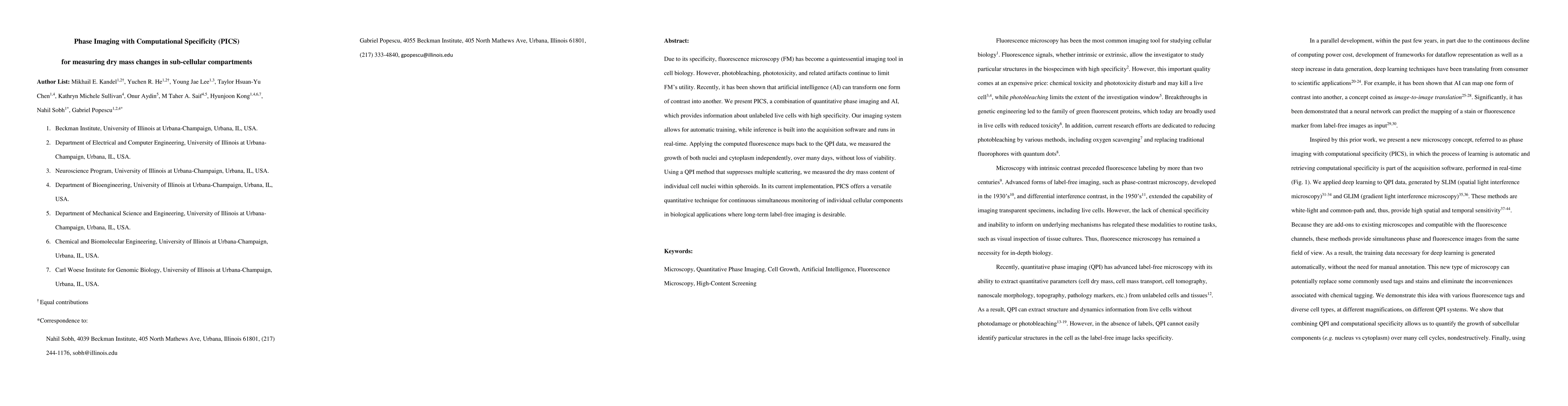
Due to its specificity, fluorescence microscopy (FM) has become a quintessential imaging tool in cell biology. However, photobleaching, phototoxicity, and related artifacts continue to limit FM's utility. Recently, it has been shown that artificial intelligence (AI) can transform one form of contrast into another. We present PICS, a combination of quantitative phase imaging and AI, which provides information about unlabeled live cells with high specificity. Our imaging system allows for automatic training, while inference is built into the acquisition software and runs in real-time. Applying the computed fluorescence maps back to the QPI data, we measured the growth of both nuclei and cytoplasm independently, over many days, without loss of viability. Using a QPI method that suppresses multiple scattering, we measured the dry mass content of individual cell nuclei within spheroids. In its current implementation, PICS offers a versatile quantitative technique for continuous simultaneous monitoring of individual cellular components in biological applications where long-term label-free imaging is desirable.
AI Key Findings
Get AI-generated insights about this paper's methodology, results, significance, and more — seven facets brought into focus.
Impact
Paper Details
PDF Preview
Key Terms
Citation Network
Current paper (gray), citations (green), references (blue)
Display is limited for performance on very large graphs.
Discussion 0