Summary
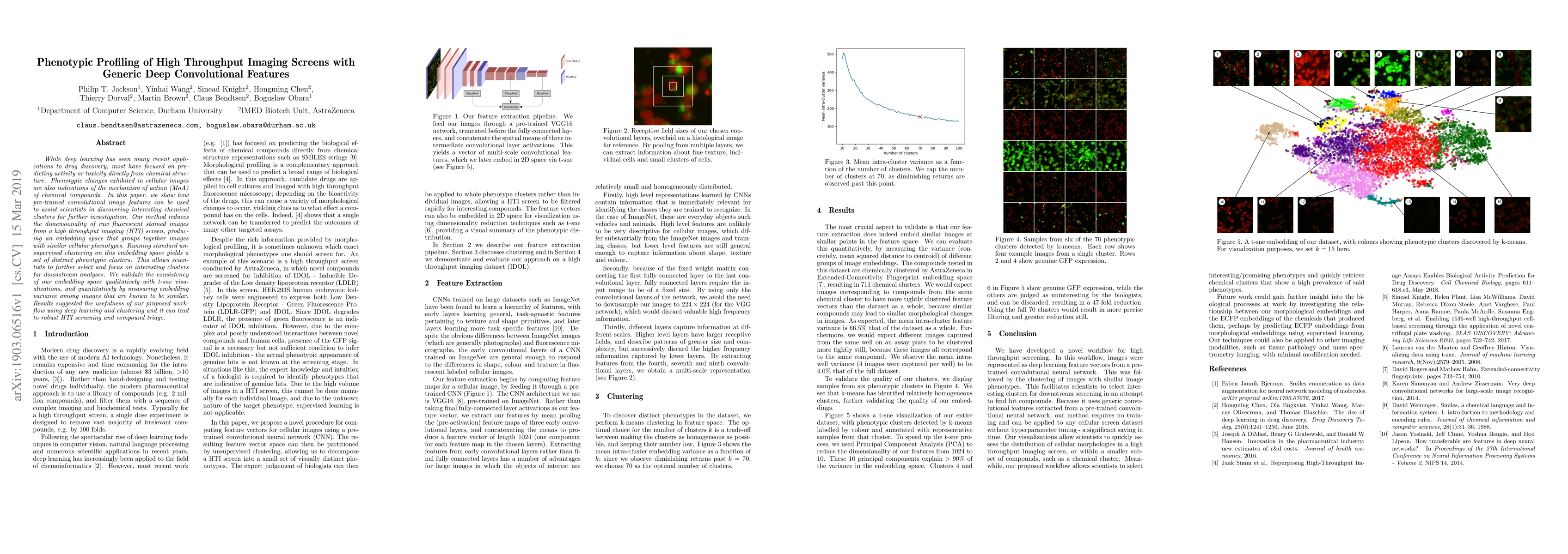
While deep learning has seen many recent applications to drug discovery, most have focused on predicting activity or toxicity directly from chemical structure. Phenotypic changes exhibited in cellular images are also indications of the mechanism of action (MoA) of chemical compounds. In this paper, we show how pre-trained convolutional image features can be used to assist scientists in discovering interesting chemical clusters for further investigation. Our method reduces the dimensionality of raw fluorescent stained images from a high throughput imaging (HTI) screen, producing an embedding space that groups together images with similar cellular phenotypes. Running standard unsupervised clustering on this embedding space yields a set of distinct phenotypic clusters. This allows scientists to further select and focus on interesting clusters for downstream analyses. We validate the consistency of our embedding space qualitatively with t-sne visualizations, and quantitatively by measuring embedding variance among images that are known to be similar. Results suggested the usefulness of our proposed workflow using deep learning and clustering and it can lead to robust HTI screening and compound triage.
AI Key Findings
Get AI-generated insights about this paper's methodology, results, and significance.
Paper Details
PDF Preview
Key Terms
Citation Network
Current paper (gray), citations (green), references (blue)
Display is limited for performance on very large graphs.
Similar Papers
Found 4 papers| Title | Authors | Year | Actions |
|---|
Comments (0)