ArxivLens

Quick Summary:

This paper introduces a photonic chip-assisted correlative light and electron microscopy (CLEM) technique that combines the versatility of light microscopy with the high resolution of electron microscopy. The photonic chip enables multi-modal total internal reflection fluorescence microscopy over large fields of view and facilitates high-precision localization for electron microscopy, demonstrating its potential for high-throughput CLEM and insights into intracellular communication.

arXiv Id: 1911.07055
Comments: 23 Pages, 11 Figures
Date Published: 2019-11-19
Date Updated: 2019-11-19

Key Terms (TF-IDF):

microscopy (0.258)
photonic (0.242)
chip (0.229)
em (0.217)
high precision (0.206)
lm (0.199)
precision (0.164)
cells (0.155)
electron (0.142)
structural (0.139)
area (0.130)
view (0.130)
layer (0.120)
high (0.118)
resolution (0.113)
sample (0.108)
fluorescence (0.107)
morphological (0.106)
waveguide (0.106)
light (0.105)
License: http://arxiv.org/licenses/nonexclusive-distrib/1.0/

Summary:

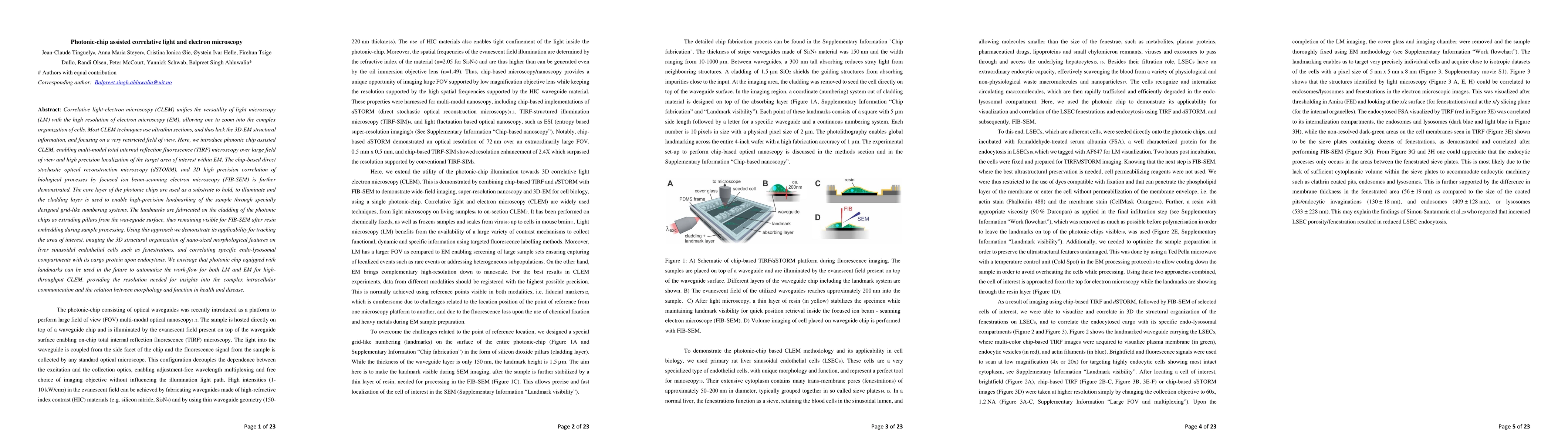
Correlative light-electron microscopy (CLEM) unifies the versatility of light microscopy (LM) with the high resolution of electron microscopy (EM), allowing one to zoom into the complex organization of cells. Most CLEM techniques use ultrathin sections, and thus lack the 3D-EM structural information, and focusing on a very restricted field of view. Here, we introduce photonic chip assisted CLEM, enabling multi-modal total internal reflection fluorescence (TIRF) microscopy over large field of view and high precision localization of the target area of interest within EM. The chip-based direct stochastic optical reconstruction microscopy (dSTORM), and 3D high precision correlation of biological processes by focused ion beam-scanning electron microscopy (FIB-SEM) is further demonstrated. The core layer of the photonic chips are used as a substrate to hold, to illuminate and the cladding layer is used to enable high-precision landmarking of the sample through specially designed grid-like numbering systems. The landmarks are fabricated on the cladding of the photonic chips as extruding pillars from the waveguide surface, thus remaining visible for FIB-SEM after resin embedding during sample processing. Using this approach we demonstrate its applicability for tracking the area of interest, imaging the 3D structural organization of nano-sized morphological features on liver sinusoidal endothelial cells such as fenestrations, and correlating specific endo-lysosomal compartments with its cargo protein upon endocytosis. We envisage that photonic chip equipped with landmarks can be used in the future to automatize the work-flow for both LM and EM for high-throughput CLEM, providing the resolution needed for insights into the complex intracellular communication and the relation between morphology and function in health and disease.

Citation & Reference Network Visualization

This graph shows how this paper is connected to other research. The current paper is shown in dark gray, papers that cite it are in green, and papers it references are in blue. You can:

Current Paper
Papers citing this work (Citations)
Papers cited by this work (References)

• Hover over any node to see the paper title

• Click a node to view that paper's details

• Drag nodes to rearrange the graph

• Use the zoom controls (bottom right) to zoom in/out

Citations and References

Title Authors Year Actions
Correlative Super‐Resolution Imaging of Cellular Nanopores Facilitated by Transparent Polymer Waveguide Chips
S. B. Dutta, Jasmin Celine Schürstedt‐Seher, Anders Kokkvoll Engdahl, Wolfgang Hübner, Stefan Belle, Karolina Szafranska, Peter McCourt, Ralf Hellmann, M. Schüttpelz, Thomas Huser
2024
View Paper
Super-resolution histology of paraffin-embedded samples via photonic chip-based microscopy
Luis E. Villegas-Hernández, Vishesh K. Dubey, H. Mao, M. Pradhan, Jean-Claude Tinguely, D. H. Hansen, Sebastián Acuña, B. Zapotoczny, Krishna Agarwal, Mona Nystad, Ganesh Acharya, K. Fenton, H. Danielsen, B. Ahluwalia
2023
View Paper
UV photonic integrated circuits for far-field structured illumination autofluorescence microscopy
Chupao Lin, Juan Santo-Domingo Peñaranda, J. Dendooven, C. Detavernier, D. Schaubroeck, N. Boon, Roel Baets, N. Le Thomas
2022
View Paper
Extracellular vesicles through the blood–brain barrier: a review
H. Ramos-Zaldívar, Iva Polakovičová, E. Salas-Huenuleo, A. Corvalán, M. Kogan, Claudia P. Yefi, M. Andía
2022
View Paper
Fluorescence CLEM in biology: historic developments and current super‐resolution applications
K. van den Dries, Jack Fransen, A. Cambi
2022
View Paper
From fixed-dried to wet-fixed to live – comparative super-resolution microscopy of liver sinusoidal endothelial cell fenestrations
K. Szafranska, Tanja Neuman, Zbigniew Baster, Z. Rajfur, O. Szelest, C. Holte, Agata Kubisiak, E. Kuś, D. Wolfson, S. Chłopicki, B. Ahluwalia, M. Lekka, M. Szymoński, P. Mccourt, B. Zapotoczny
2022
View Paper
Chip-based multimodal super-resolution microscopy for histological investigations of cryopreserved tissue sections
Luis E. Villegas-Hernández, Vishesh K. Dubey, Mona Nystad, Jean-Claude Tinguely, D. Coucheron, F. Dullo, A. Priyadarshi, Sebastián Acuña, Azeem Ahmad, J. Mateos, Gery Barmettler, U. Ziegler, Å. Birgisdottir, A. K. Hovd, K. Fenton, Ganesh Acharya, Krishna Agarwal, B. Ahluwalia
2022
View Paper
Photonic chip-based multimodal super-resolution microscopy for histopathological assessment of cryopreserved tissue sections
Luis E. Villegas-Hernández, Vishesh K. Dubey, Mona Nystad, Jean-Claude Tinguely, D. Coucheron, F. Dullo, A. Priyadarshi, Sebastián Acuña, J. Mateos, Gery Barmettler, U. Ziegler, A. K. Hovd, K. Fenton, G. Acharya, Krishna Agarwal, B. Ahluwalia
2021
View Paper

Similar Papers

Guided-deconvolution for Correlative Light and Electron Microscopy

Rainer Heintzmann, Fengjiao Ma, Rainer Kaufmann, Jaroslaw Sedzicki, Zoltán Cseresnyés, Christoph Dehio, Stephanie Hoeppener, Marc Thilo Figge

Published: 2022-08-22

Correlative light electron microscopy using small gold nanoparticles as single probes

Wolfgang Langbein, Lukas Payne, Paola Borri, Iestyn Pope, Hugh Tanner, Francesco Masia, Kenton Paul Arkill, Judith Mantell, Paul Verkade

Published: 2023-12-05

Comments (0)

This website uses cookies to ensure you get the best experience. Learn more