This paper proposes a 
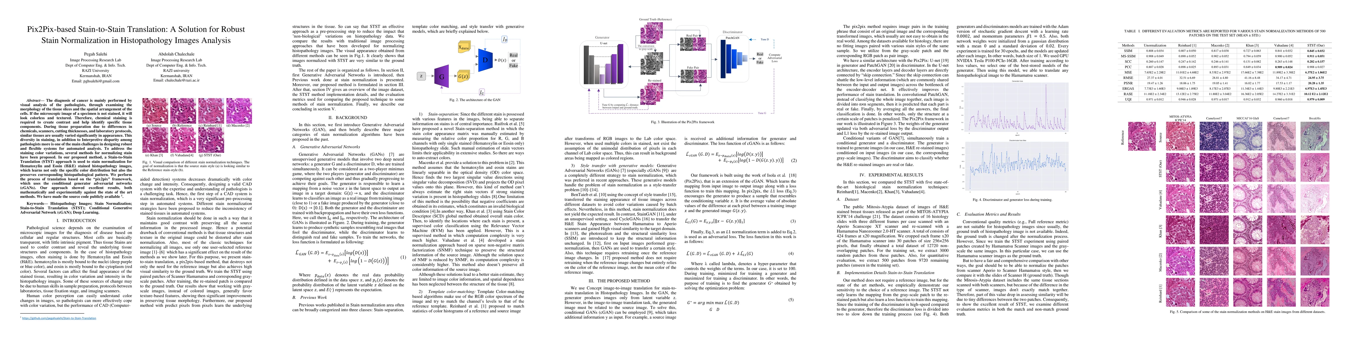
Pix2Pix-based Stain-to-Stain Translation (STST) method for normalizing color variations in Hematoxylin and Eosin (H&E) stained histopathology images, using conditional GANs to maintain histopathological patterns. The method outperforms existing techniques in both mathematical and experimental evaluations.
The diagnosis of cancer is mainly performed by visual analysis of the pathologists, through examining the morphology of the tissue slices and the spatial arrangement of the cells. If the microscopic image of a specimen is not stained, it will look colorless and textured. Therefore, chemical staining is required to create contrast and help identify specific tissue components. During tissue preparation due to differences in chemicals, scanners, cutting thicknesses, and laboratory protocols, similar tissues are usually varied significantly in appearance. This diversity in staining, in addition to Interpretive disparity among pathologists more is one of the main challenges in designing robust and flexible systems for automated analysis. To address the staining color variations, several methods for normalizing stain have been proposed. In our proposed method, a Stain-to-Stain Translation (STST) approach is used to stain normalization for Hematoxylin and Eosin (H&E) stained histopathology images, which learns not only the specific color distribution but also the preserves corresponding histopathological pattern. We perform the process of translation based on the pix2pix framework, which uses the conditional generator adversarial networks (cGANs). Our approach showed excellent results, both mathematically and experimentally against the state of the art methods. We have made the source code publicly available.
This graph shows how this paper is connected to other research. The current paper is shown in dark gray, papers that cite it are in green, and papers it references are in blue. You can:
• Hover over any node to see the paper title
• Click a node to view that paper's details
• Drag nodes to rearrange the graph
• Use the zoom controls (bottom right) to zoom in/out
Title | Authors | Year | Actions |
---|---|---|---|
Learnable color space conversion and fusion for stain normalization in pathology images
|
Jing Ke, Yijin Zhou, Yiqing Shen, Yi Guo, Ning Liu, Xiaodan Han, Dinggang Shen
|
2024
|
View Paper |
Enhancing GANs with Contrastive Learning-Based Multistage Progressive Finetuning SNN and RL-Based External Optimization
|
Osama Mustafa
|
2024
|
View Paper |
Stain Normalization of Hematology Slides using Neural Color Transfer
|
M. M. Arshad, Hasan Sajid, M. J. Khan
|
2024
|
View Paper |
Enhanced U-Net Framework for Nuclei Segmentation in Triple Negative Breast Cancer
|
Sushma Nagdeote, Sapna U. Prabhu
|
2024
|
View Paper |
Dataset Distillation for Histopathology Image Classification
|
Cong Cong, Shi Xuan, Sidong Liu, M. Pagnucco, Shiliang Zhang, Yang Song
|
2024
|
View Paper |
Computational methods and biomarker discovery strategies for spatial proteomics: a review in immuno-oncology
|
Haoyang Mi, Shamilene Sivagnanam, W. Ho, Shuming Zhang, Daniel Bergman, Atul Deshpande, Alexander S. Baras, Elizabeth M. Jaffee, L. Coussens, E. Fertig, Aleksander S. Popel
|
2024
|
View Paper |
Generative models for color normalization in digital pathology and dermatology: Advancing the learning paradigm
|
Massimo Salvi, Francesco Branciforti, Filippo Molinari, K. Meiburger
|
2024
|
View Paper |
StainSWIN: Vision transformer-based stain normalization for histopathology image analysis
|
Elif Baykal Kablan, Selen Ayas
|
2024
|
View Paper |
StampOne: Addressing Frequency Balance in Printer-proof Steganography
|
Farhad Shadmand, Iurii Medvedev, Luiz Schirmer, João Marcos, Nuno Gonçalves
|
2024
|
View Paper |
Wavelet Knowledge Distillation for Efficient Stain Normalization in Digital Histopathology
|
Sefa Keklik, Murat Ekinci, Elif Baykal Kablan
|
2024
|
View Paper |
Investigating staining variance effects on deep learning-based semantic segmentation in digital pathology
|
Amine Marzouki, Zografoula Vagena, Camille Kurtz, N. Loménie
|
2024
|
View Paper |
StainFuser: Controlling Diffusion for Faster Neural Style Transfer in Multi-Gigapixel Histology Images
|
R. Jewsbury, Ruoyu Wang, A. Bhalerao, N. Rajpoot, Q. Vu
|
2024
|
View Paper |
Unsupervised domain adaptation for histopathology image segmentation with incomplete labels
|
Huihui Zhou, Yan Wang, Benyan Zhang, Chunhua Zhou, Maxim S. Vonsky, L. Mitrofanova, Duowu Zou, Qingli Li
|
2024
|
View Paper |
Analyzing Generative Models for Realistic Data Augmentation across Modalities and Applications
|
Sanskruti Patel, Unnati A. Patel, Jay Nanavati, Atul Patel
|
2024
|
View Paper |
BCD-net: Stain separation of histological images using deep variational Bayesian blind color deconvolution
|
Shuowen Yang, Fernando Pérez-Bueno, Francisco M. Castro-Macías, R. Molina, A. Katsaggelos
|
2024
|
View Paper |
A New Method of Artificial-Intelligence-Based Automatic Identification of Lymphovascular Invasion in Urothelial Carcinomas
|
Bogdan Ceachi, Mirela Cioplea, P. Mustățea, Julian Gerald Dcruz, Sabina Zurac, Victor Cauni, C. Popp, Cristian Mogodici, Liana Sticlaru, A. Cioroianu, Mihai Busca, Oana Stefan, Irina Tudor, Carmen Dumitru, Alexandra Vilaia, Alexandra Oprisan, Alexandra Bastian, L. Nichita
|
2024
|
View Paper |
Chrometect GAYO: Classification and Colorization using PIX2PIX and YOLOV8
|
Nithya Madhasu, S. Pande
|
2023
|
View Paper |
A deep learning model to predict Ki-67 positivity in oral squamous cell carcinoma
|
Francesco Martino, G. Ilardi, S. Varricchio, D. Russo, R. D. Di Crescenzo, Stefania Staibano, F. Merolla
|
2023
|
View Paper |
Improving performance in colorectal cancer histology decomposition using deep and ensemble machine learning
|
Fabi Prezja, L. Annala, Sampsa Kiiskinen, Suvi Lahtinen, Timo Ojala, Pekka Ruusuvuori, Teijo Kuopio
|
2023
|
View Paper |
Segmentation of Nuclei in H&E-Stained Histological Images using Deep Learning Framework: A Perspective on Ensemble Approach and Nuclei Count
|
Madhavi Aghera, Krishna Veer Singh, Kishankumar Vaishnani, Urvi Oza, Bakul Gohel
|
2023
|
View Paper |
Class-Specific Data Augmentation: Bridging the Imbalance in Multiclass Breast Cancer Classification
|
Kanan Mahammadli, Abdullah Burkan Bereketoglu, A. Kabakcı
|
2023
|
View Paper |
Digital Staining of White Blood Cells With Confidence Estimation
|
A. Tomczak, Slobodan Ilic, Gaby Marquardt, Thomas Engel, N. Navab, Shadi Albarqouni
|
2023
|
View Paper |
Stain normalization methods for histopathology image analysis: A comprehensive review and experimental comparison
|
Md. Ziaul Hoque, A. Keskinarkaus, Pia Nyberg, T. Seppänen
|
2023
|
View Paper |
Generative Adversarial Networks for Stain Normalisation in Histopathology
|
Jack Breen, K. Zucker, Katie Allen, N. Ravikumar, N. Orsi
|
2023
|
View Paper |
Assessment of Color Reproducibility and Mitigation of Color Variation in Whole Slide Image Scanners for Toxicologic Pathology
|
Mei-Lan Chu, Xing-Yue M Ge, Jeffrey Eastham, Trung Nguyen, R. Fuji, R. Sullivan, Daniel Ruderman
|
2023
|
View Paper |
A stain color normalization with robust dictionary learning for breast cancer histological images processing
|
T. A. Tosta, A. D. Freitas, P. D. Faria, L. A. Neves, A. S. Martins, M. Z. Nascimento
|
2023
|
View Paper |
Self-Supervised Digital Histopathology Image Disentanglement for Arbitrary Domain Stain Transfer
|
Yu Ling, Weimin Tan, Bo Yan
|
2023
|
View Paper |
A Comprehensive Review on Breast Cancer Detection, Classification and Segmentation Using Deep Learning
|
Barsha Abhisheka, S. Biswas, B. Purkayastha
|
2023
|
View Paper |
Stain Transfer using CycleGAN for Histopathological Images
|
Lorenzo Veronese, Isabella Poles, E. D’Arnese, M. Santambrogio
|
2023
|
View Paper |
ContriMix: Scalable stain color augmentation for domain generalization without domain labels in digital pathology
|
Tan H. Nguyen, Dinkar Juyal, Jin Li, A. Prakash, Shima Nofallah, Chintan Shah, Sai Chowdary Gullapally, Limin Yu, Michael Griffin, Anand Sampat, J. Abel, Justin Lee, A. Taylor-Weiner
|
2023
|
View Paper |
Regional realness-aware generative adversarial networks for stain normalization
|
Elif Baykal Kablan
|
2023
|
View Paper |
A Medically Assisted Model for Precise Segmentation of Osteosarcoma Nuclei on Pathological Images
|
Jia Wu, Tingyu Yuan, Jiachen Zeng, Fangfang Gou
|
2023
|
View Paper |
ParamNet: A Dynamic Parameter Network for Fast Multi-to-One Stain Normalization
|
Hongtao Kang, Die Luo, Li Chen, Junbo Hu, Tingwei Quan, Shaoqun Zeng, Shenghua Cheng, Xiuli Liu
|
2023
|
View Paper |
CDA-Net: A contrastive deep adversarial model for prostate cancer segmentation in MRI images
|
Zhixun Li, Jiancheng Fang, Ruiyun Qiu, Huiling Gong, Wei Zhang, Linghao Li, Jian-Jun Jiang
|
2023
|
View Paper |
The promise and challenge of spatial omics in dissecting tumour microenvironment and the role of AI
|
R. Y. Lee, Chan Way Ng, Menaka Rajapakse, N. Ang, J. Yeong, M. Lau
|
2023
|
View Paper |
Automatic normalized digital color staining in the recognition of abnormal blood cells using generative adversarial networks
|
Kevin Barrera, J. Rodellar, Santiago Alférez, A. Merino
|
2023
|
View Paper |
Ink removal in whole slide images using hallucinated data
|
V. Ramanathan, Wenchao Han, Dina Bassiouny, E. Rakovitch, Anne L. Martel
|
2023
|
View Paper |
A progressive phased attention model fused histopathology image features and gene features for lung cancer staging prediction
|
Meiling Cai, Lin Zhao, Yanan Zhang, Wei Wu, Liye Jia, Juanjuan Zhao, Qianqian Yang, Yan Qiang
|
2023
|
View Paper |
Red-green-blue to normalized difference vegetation index translation: a robust and inexpensive approach for vegetation monitoring using machine vision and generative adversarial networks
|
A. Farooque, Hassan Afzaal, Rachid Benlamri, Salem Al-Naemi, Evan MacDonald, F. Abbas, Kaelyn MacLeod, H. Ali
|
2023
|
View Paper |
Impact of Stain Normalization on Pathologist Assessment of Prostate Cancer: A Comparative Study
|
M. Salvi, Alessandro Caputo, D. Balmativola, Manuela Scotto, Orazio Pennisi, Nicola Michielli, Alessandro Mogetta, F. Molinari, F. Fraggetta
|
2023
|
View Paper |
Impact of Stain Normalisation Technique on Deep Learning based Nuclei Segmentation in Histopathological Image
|
Kishankumar Vaishnani, Bakul Gohel, Avik Hati
|
2023
|
View Paper |
Thermal image generation for blast furnace chute based on generative adversarial network
|
Xiaoman Cheng, S. Cheng
|
2023
|
View Paper |
Multi-domain stain normalization for digital pathology: A cycle-consistent adversarial network for whole slide images
|
M. J. Hetz, Tabea-Clara Bucher, T. Brinker
|
2023
|
View Paper |
Stain normalization using score-based diffusion model through stain separation and overlapped moving window patch strategies
|
Jiheon Jeong, Ki Duk Kim, Yujin Nam, C. Cho, H. Go, Namkug Kim
|
2023
|
View Paper |
A Fast Texture-to-Stain Adversarial Stain Normalization Network for Histopathological Images
|
Qi Jia, Jing Guo, Fei Du, Peng Yang, Yun Yang
|
2022
|
View Paper |
A Federated Learning System for Histopathology Image Analysis With an Orchestral Stain-Normalization GAN
|
Yiqing Shen, A. Sowmya, Yulin Luo, Xiaoyao Liang, Dinggang Shen, Jing Ke
|
2022
|
View Paper |
Deep learning-enabled virtual histological staining of biological samples
|
Bijie Bai, Xilin Yang, Yuzhu Li, Yijie Zhang, N. Pillar, Aydogan Ozcan
|
2022
|
View Paper |
HistoStarGAN: A Unified Approach to Stain Normalisation, Stain Transfer and Stain Invariant Segmentation in Renal Histopathology
|
Jelica Vasiljevi'c, F. Feuerhake, Cédric Wemmert, T. Lampert
|
2022
|
View Paper |
Comparative analysis of segmentation and generative models for fingerprint retrieval task
|
Megha Patel, Devarsh Patel, Sarthak Patel
|
2022
|
View Paper |
Stain normalization in digital pathology: Clinical multi-center evaluation of image quality
|
Nicola Michielli, Alessandro Caputo, Manuela Scotto, Alessandro Mogetta, Orazio Pennisi, F. Molinari, D. Balmativola, M. Bosco, A. Gambella, J. Metovic, Daniele Tota, L. Carpenito, P. Gasparri, M. Salvi
|
2022
|
View Paper |
Tackling stain variability using CycleGAN-based stain augmentation
|
Nassim Bouteldja, David L. Hölscher, R. D. Bülow, I. S. Roberts, Rosanna Coppo, P. Boor
|
2022
|
View Paper |
DermoCC-GAN: A new approach for standardizing dermatological images using generative adversarial networks
|
M. Salvi, Francesco Branciforti, F. Veronese, E. Zavattaro, V. Tarantino, P. Savoia, K. Meiburger
|
2022
|
View Paper |
Impact of scanner variability on lymph node segmentation in computational pathology
|
Amjad Khan, A. Janowczyk, Felix Müller, A. Blank, Huu-Giao Nguyen, C. Abbet, Linda Studer, A. Lugli, H. Dawson, J. Thiran, I. Zlobec
|
2022
|
View Paper |
RandStainNA: Learning Stain-Agnostic Features from Histology Slides by Bridging Stain Augmentation and Normalization
|
Yiqing Shen, Yulin Luo, Dinggang Shen, Jing Ke
|
2022
|
View Paper |
A SSIM Guided cGAN Architecture For Clinically Driven Generative Image Synthesis of Multiplexed Spatial Proteomics Channels
|
Jillur Rahman Saurav, M. Nasr, Paul Koomey, Michael Robben, Manfred Huber, J. Weidanz, B. Ryan, E. Ruppin, Peng-Tao Jiang, Jacob M. Luber
|
2022
|
View Paper |
RestainNet: a self-supervised digital re-stainer for stain normalization
|
Bingchao Zhao, Jiatai Lin, C. Liang, Zongjian Yi, Xin Chen, Bingbing Li, Weihao Qiu, Dan Li, Li Liang, Chu Han, Zaiyi Liu
|
2022
|
View Paper |
High-throughput widefield fluorescence imaging of 3D samples using deep learning for 2D projection image restoration
|
Edvin Forsgren, Christoffer Edlund, Miniver Oliver, Kalpana Barnes, Rickard Sjögren, Timothy R. Jackson
|
2022
|
View Paper |
Bayesian K-SVD for H and E blind color deconvolution. Applications to stain normalization, data augmentation and cancer classification
|
Fernando Pérez-Bueno, J. G. Serra, M. Vega, J. Mateos, R. Molina, A. Katsaggelos
|
2022
|
View Paper |
Inter-Semantic Domain Adversarial in Histopathological Images
|
Nicolas Dumas, V. Derangère, L. Arnould, S. Ladoire, Louis-Oscar Morel, Nathan Vinçon
|
2022
|
View Paper |
Improving unsupervised stain-to-stain translation using self-supervision and meta-learning
|
Nassim Bouteldja, B. Klinkhammer, Tarek Schlaich, P. Boor, D. Merhof
|
2021
|
View Paper |
Evaluation of normalization technique on classification with deep learning features
|
A. D. Freitas, Adriano B. Silva, A. S. Martins, L. A. Neves, T. A. Tosta, P. D. Faria, M. Z. Nascimento
|
2021
|
View Paper |
Deep learning 2D and 3D optical sectioning microscopy using cross-modality Pix2Pix cGAN image translation.
|
Huimin Zhuge, B. Summa, Jihun Hamm, J. Q. Brown
|
2021
|
View Paper |
Generative Adversarial Networks in Digital Pathology and Histopathological Image Processing: A Review
|
Laya Jose, Sidong Liu, C. Russo, A. Nadort, A. Di Ieva
|
2021
|
View Paper |
The utility of color normalization for AI‐based diagnosis of hematoxylin and eosin‐stained pathology images
|
J. Boschman, H. Farahani, Amirali Darbandsari, P. Ahmadvand, Ashley Van Spankeren, D. Farnell, Adrian B Levine, J. Naso, A. Churg, Steven J. M. Jones, Stephen S. F. Yip, M. Koebel, D. Huntsman, B. Gilks, Ali Bashashati
|
2021
|
View Paper |
Generation of the NIR Spectral Band for Satellite Images with Convolutional Neural Networks
|
S. Illarionova, Dmitrii G. Shadrin, A. Trekin, V. Ignatiev, I. Oseledets
|
2021
|
View Paper |
Texture Enhanced Generative Adversarial Network For Stain Normalisation In Histopathology Images
|
Cong Cong, Sidong Liu, A. Ieva, M. Pagnucco, S. Berkovsky, Yang Song
|
2021
|
View Paper |
GENERATIVE DEEP LEARNING IN DIGITAL PATHOLOGY WORKFLOWS.
|
David Morrison, David Harris-Birtill, P. Caie
|
2021
|
View Paper |
StainNet: A Fast and Robust Stain Normalization Network
|
Hongtao Kang, Die Luo, Weihua Feng, Li Chen, Junbo Hu, Shaoqun Zeng, Tingwei Quan, Xiuli Liu
|
2020
|
View Paper |
Generative Adversarial Networks (GANs): An Overview of Theoretical Model, Evaluation Metrics, and Recent Developments
|
Pegah Salehi, A. Chalechale, M. Taghizadeh
|
2020
|
View Paper |
Contactless Palmprint Image Recognition Across Smartphones With Self-Paced CycleGAN
|
Qi Zhu, Guangnan Xin, Lunke Fei, Dong Liang, Zheng Zhang, Daoqiang Zhang, Dafan Zhang
|
2023
|
View Paper |
ContriMix: Unsupervised disentanglement of content and attribute for domain generalization in microscopy image analysis
|
Tan H. Nguyen, Dinkar Juyal, Jin Li, Aaditya (Adi) Prakash, Shima Nofallah, Chintan Shah, Sai Chowdary Gullapally, Michael Griffin, Anand Sampat, J. Abel, Justin Lee, A. Taylor-Weiner
|
2023
|
View Paper |
Progress of machine vision in the detection of cancer cells in histopathology
|
Wenbin He, Yongjie Han, Wuyi Ming, Jinguang Du, Yinxia Liu, Yuan Yang, Leijie Wang, Yongqiang Wang, Zhiwen Jiang, Chen Cao, Jie Yuan
|
2022
|
View Paper |
The devil is in the details: Whole Slide Image acquisition and processing for artifacts detection, color variation, and data augmentation. A review.
|
Neel Kanwal, Fernando Pérez-Bueno, Arne Schmidt, K. Engan, R. Molina
|
2022
|
View Paper |
Whole Slide Image Quality in Digital Pathology: Review and Perspectives
|
Romain Brixtel, Sébastien Bougleux, O. Lézoray, Yann Caillot, Benoît Lemoine, Mathieu Fontaine, Dalal Nebati, Arnaud Renouf
|
2022
|
View Paper |
Semi-supervised Adversarial Learning for Stain Normalisation in Histopathology Images
|
Cong Cong, Sidong Liu, A. Ieva, M. Pagnucco, S. Berkovsky, Yang Song
|
2021
|
View Paper |
Master Thesis-Medical Image Analysis using Deep Learning
|
Stephan, Declaration OF Originality
|
2020
|
View Paper |
Medical Image Analysis
|
Hongtao Kang, Li Chen, Junbo Hu, Tingwei Quan, Shaoqun Zeng, Shenghua Cheng, Xiuli Liu
|
View Paper |
Nicolas Brieu, Philipp Wortmann, Ansh Kapil, Felix J. Segerer, Guenter Schmidt
Published: 2022-07-04
Published: 2021-06-04
Dinggang Shen, Yiqing Shen, Jing Ke, Yulin Luo
Published: 2023-03-03
Comments (0)