Summary
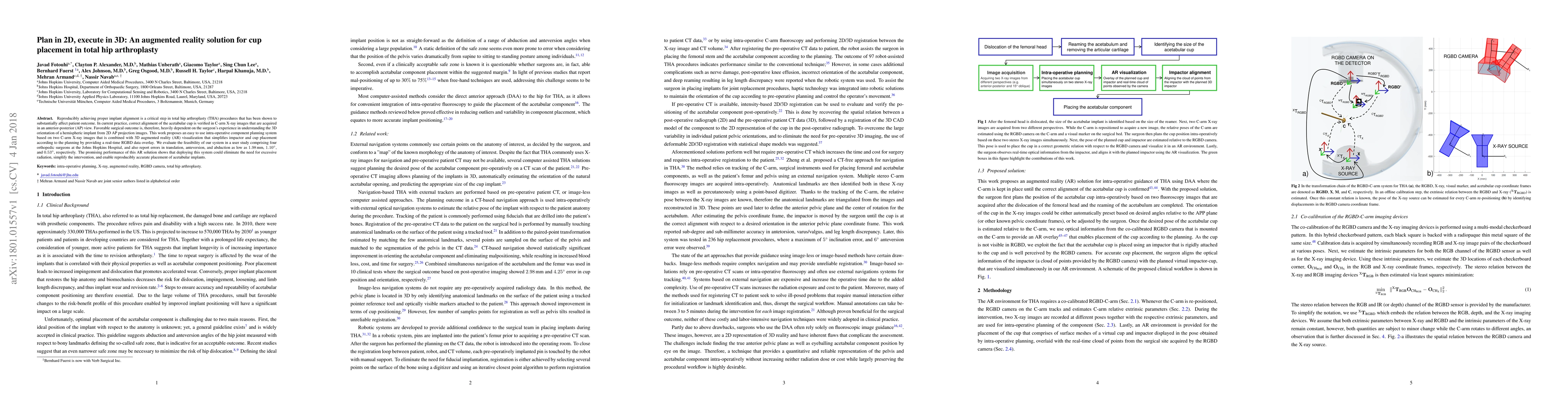
Reproducibly achieving proper implant alignment is a critical step in total hip arthroplasty (THA) procedures that has been shown to substantially affect patient outcome. In current practice, correct alignment of the acetabular cup is verified in C-arm X-ray images that are acquired in an anterior-posterior (AP) view. Favorable surgical outcome is, therefore, heavily dependent on the surgeon's experience in understanding the 3D orientation of a hemispheric implant from 2D AP projection images. This work proposes an easy to use intra-operative component planning system based on two C-arm X-ray images that is combined with 3D augmented reality (AR) visualization that simplifies impactor and cup placement according to the planning by providing a real-time RGBD data overlay. We evaluate the feasibility of our system in a user study comprising four orthopedic surgeons at the Johns Hopkins Hospital, and also report errors in translation, anteversion, and abduction as low as 1.98 mm, 1.10 degrees, and 0.53 degrees, respectively. The promising performance of this AR solution shows that deploying this system could eliminate the need for excessive radiation, simplify the intervention, and enable reproducibly accurate placement of acetabular implants.
AI Key Findings
Generated Sep 02, 2025
Methodology
The research proposes an augmented reality (AR) solution for cup placement in total hip arthroplasty (THA) using two C-arm X-ray images combined with 3D AR visualization, simplifying impactor and cup placement in real-time.
Key Results
- The system achieved translation, anteversion, and abduction errors as low as 1.98 mm, 1.10 degrees, and 0.53 degrees, respectively.
- The AR solution minimized anteversion and abduction errors substantially compared to the classic DAA approach.
- All surgeons participating in the user study found presetting the cup orientation useful and valid.
Significance
This AR solution has the potential to reduce radiation exposure, operating time, and frustration while increasing efficiency and accuracy in placing acetabular components, ultimately aiding in reducing the risk of revision surgery in patients with diseased hip joints.
Technical Contribution
The development of an AR environment built upon an RGB-D-enhanced C-arm, enabling visualization of 3D optical information from the surgical site superimposed with the planning target.
Novelty
This work stands out by proposing an AR solution for THA that requires no pre-operative data, is performed only on two stereo X-ray images, and reduces the surgeon's task load by automatically adjusting the cup's orientation if one of the X-ray projections is from an AP perspective.
Limitations
- The study did not discuss any specific limitations.
- Further research and user studies on cadaveric specimens are needed to quantify changes in operating time, number of required X-ray images, dose, accuracy, and surgical task load compared to classic image-guided approaches.
Future Work
- Investigate the use of pose-aware RGB-D augmented C-arm for acquiring and confirming true AP images considering pelvis tilts in different planes.
- Explore the incorporation of RGB-D-based simultaneous localization and mapping to track the surgical site without external visual markers.
Paper Details
PDF Preview
Key Terms
Citation Network
Current paper (gray), citations (green), references (blue)
Display is limited for performance on very large graphs.
Similar Papers
Found 4 papersInvestigation of Cup Placement Position in Total Hip Arthroplasty with Cup-side Implant Placement in Computed Tomography Horizontal Sections.
Furuichi, Shuro, Mitani, Shigeru, Endo, Hirosuke et al.
| Title | Authors | Year | Actions |
|---|
Comments (0)