Planar 3D Transfer Learning for End to End Unimodal MRI Unbalanced Data Segmentation
Publication
Metrics
AI Quick Summary
This paper presents a novel 2D to 3D transfer learning method for segmenting unbalanced MRI data using a planar 3D res-u-net network, achieving high sensitivity and Dice score. The approach uses pre-trained 2D VGG-16 weights and performs well on the MICCAI 2016 MS lesion challenge dataset, demonstrating practical utility in real medical practice.
Paper Preview
Abstract
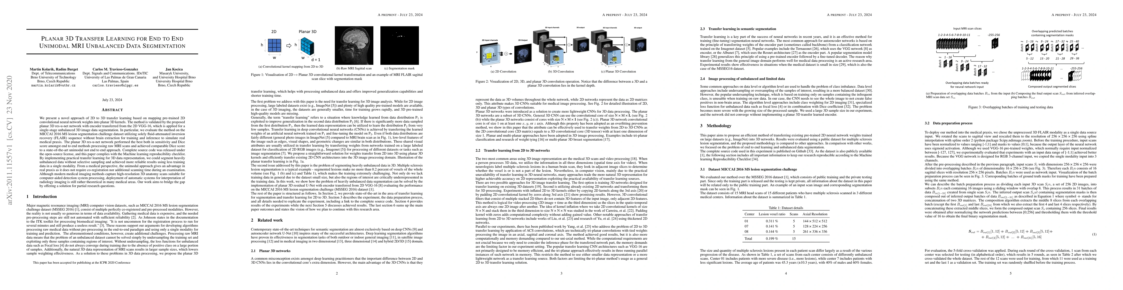
We present a novel approach of 2D to 3D transfer learning based on mapping pre-trained 2D convolutional neural network weights into planar 3D kernels. The method is validated by the proposed planar 3D res-u-net network with encoder transferred from the 2D VGG-16, which is applied for a single-stage unbalanced 3D image data segmentation. In particular, we evaluate the method on the MICCAI 2016 MS lesion segmentation challenge dataset utilizing solely fluid-attenuated inversion recovery (FLAIR) sequence without brain extraction for training and inference to simulate real medical praxis. The planar 3D res-u-net network performed the best both in sensitivity and Dice score amongst end to end methods processing raw MRI scans and achieved comparable Dice score to a state-of-the-art unimodal not end to end approach. Complete source code was released under the open-source license, and this paper complies with the Machine learning reproducibility checklist. By implementing practical transfer learning for 3D data representation, we could segment heavily unbalanced data without selective sampling and achieved more reliable results using less training data in a single modality. From a medical perspective, the unimodal approach gives an advantage in real praxis as it does not require co-registration nor additional scanning time during an examination. Although modern medical imaging methods capture high-resolution 3D anatomy scans suitable for computer-aided detection system processing, deployment of automatic systems for interpretation of radiology imaging is still rather theoretical in many medical areas. Our work aims to bridge the gap by offering a solution for partial research questions.
AI Key Findings
Get AI-generated insights about this paper's methodology, results, significance, and more — seven facets brought into focus.
Impact
Paper Details
Authors
PDF Preview
Key Terms
Citation Network
Current paper (gray), citations (green), references (blue)
Display is limited for performance on very large graphs.
Discussion 0