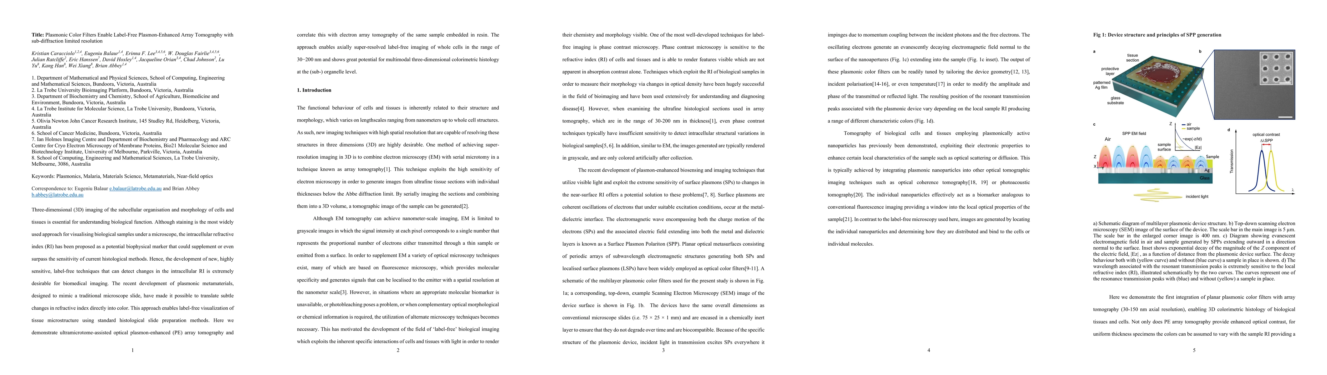
Plasmonic Color Filters Enable Label-Free Plasmon-Enhanced Array Tomography with sub-diffraction limited resolution
Publication
Metrics
Paper Preview
Abstract
Three-dimensional (3D) imaging of the subcellular organisation and morphology of cells and tissues is essential for understanding biological function. Although staining is the most widely used approach for visualising biological samples under a microscope, the intracellular refractive index (RI) has been proposed as a potential biophysical marker that could supplement or even surpass the sensitivity of current histological methods. Hence, the development of new, highly sensitive, label-free techniques that can detect changes in the intracellular RI is extremely desirable for biomedical imaging. The recent development of plasmonic metamaterials, designed to mimic a traditional microscope slide, have made it possible to translate subtle changes in refractive index directly into color. This approach enables label-free visualization of tissue microstructure using standard histological slide preparation methods. Here we demonstrate ultramicrotome-assisted optical plasmon-enhanced (PE) array tomography and correlate this with electron array tomography of the same sample embedded in resin. The approach enables axially super-resolved label-free imaging of whole cells in the range of 30-200 nm and shows great potential for multimodal three-dimensional colorimetric histology at the (sub-) organelle level.
AI Key Findings
Get AI-generated insights about this paper's methodology, results, significance, and more — seven facets brought into focus.
Impact
Paper Details
Authors
PDF Preview
Citation Network
Current paper (gray), citations (green), references (blue)
Display is limited for performance on very large graphs.
Discussion 0