Publication
Metrics
AI Quick Summary
This paper introduces an x-ray plenoptic microscope utilizing a micro-capillary array to capture multiple x-ray projections in a single exposure, enabling depth-resolved tomographic imaging of small subvolumes within large samples, and facilitating the imaging of weakly absorbing objects through phase-contrast.
Paper Preview
Abstract
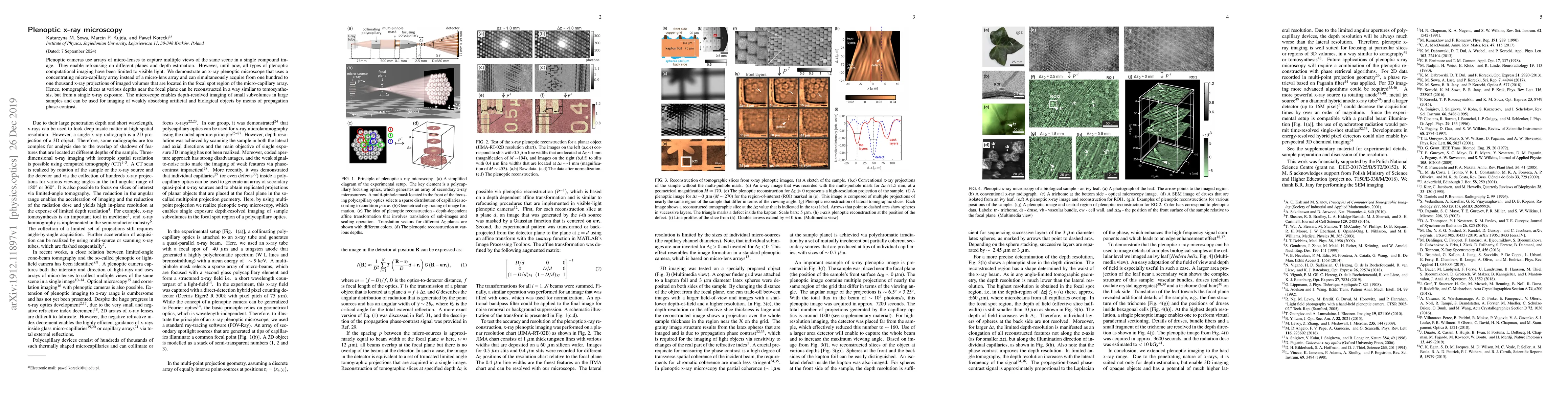
Plenoptic cameras use arrays of micro-lenses to capture multiple views of the same scene in a single compound image. They enable refocusing on different planes and depth estimation. However, until now, all types of plenoptic computational imaging have been limited to visible light. We demonstrate an x-ray plenoptic microscope that uses a concentrating micro-capillary array instead of a micro-lens array and can simultaneously acquire from one hundred to one thousand x-ray projections of imaged volumes that are located in the focal spot region of the micro-capillary array. Hence, tomographic slices at various depths near the focal plane can be reconstructed in a way similar to tomosynthesis, but from a single x-ray exposure. The microscope enables depth-resolved imaging of small subvolumes in large samples and can be used for imaging of weakly absorbing artificial and biological objects by means of propagation phase-contrast.
AI Key Findings
Get AI-generated insights about this paper's methodology, results, significance, and more — seven facets brought into focus.
Impact
Paper Details
Authors
PDF Preview
Key Terms
Citation Network
Current paper (gray), citations (green), references (blue)
Display is limited for performance on very large graphs.
Discussion 0