ArxivLens
ArXiv eess.IV cs.CV

Polyp Segmentation Using Wavelet-Based Cross-Band Integration for Enhanced Boundary Representation

Haesung Oh, Jaesung Lee

2603.03682 Published Mar 4, 2026
View PDF

Publication

Published: Mar 04, 2026
Categories: eess.IV, cs.CV

Metrics

Source: ArXiv

Quick Answers

What methodology did the authors use?

Dual-encoder Res2Net architecture that processes RGB and grayscale images separately, fuses frequency-aligned features via Band‑Specific Window Cross‑Attention (BS‑WCA) and refines them with Cascade Dilated Fusion (CDF) to enhance boundary precision. More in Methodology →

What are the key results?

Wavelet analysis shows grayscale sub‑bands consistently exhibit higher boundary contrast than RGB across all frequency bands. — The proposed model achieves higher mean Dice and IoU scores than state‑of‑the‑art baselines on Kvasir‑SEG, CVC‑ClinicDB, CVC‑ColonDB, and ETIS datasets. More in Key Results →

Why is this work significant?

By improving polyp boundary delineation, the method supports earlier and more accurate colorectal cancer detection, potentially reducing missed lesions in clinical practice. More in Significance →

What are the main limitations?

Evaluation relies solely on Dice and IoU; boundary‑sensitive metrics were not reported. — The contribution of each frequency band and modality was not isolated through ablation studies. More in Limitations →

Paper Preview

Abstract

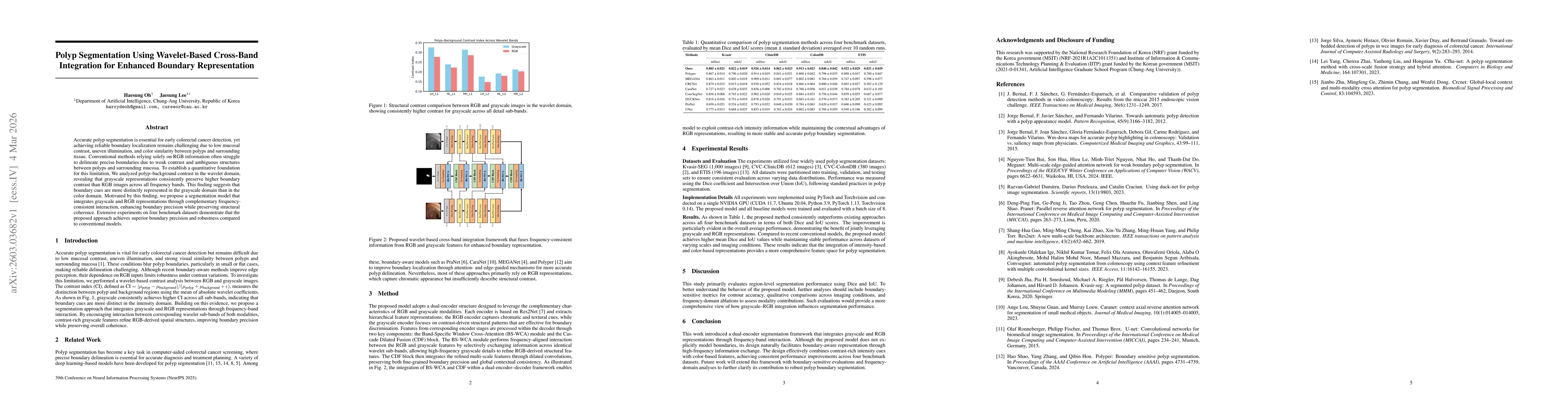
Accurate polyp segmentation is essential for early colorectal cancer detection, yet achieving reliable boundary localization remains challenging due to low mucosal contrast, uneven illumination, and color similarity between polyps and surrounding tissue. Conventional methods relying solely on RGB information often struggle to delineate precise boundaries due to weak contrast and ambiguous structures between polyps and surrounding mucosa. To establish a quantitative foundation for this limitation, we analyzed polyp-background contrast in the wavelet domain, revealing that grayscale representations consistently preserve higher boundary contrast than RGB images across all frequency bands. This finding suggests that boundary cues are more distinctly represented in the grayscale domain than in the color domain. Motivated by this finding, we propose a segmentation model that integrates grayscale and RGB representations through complementary frequency-consistent interaction, enhancing boundary precision while preserving structural coherence. Extensive experiments on four benchmark datasets demonstrate that the proposed approach achieves superior boundary precision and robustness compared to conventional models.

Key Findings, in focus

Seven facets of this paper, analysed and brought into focus by AI.

AI-generated · verify against source Generated Mar 05, 2026
In focus · one-line synthesis

By improving polyp boundary delineation, the method supports earlier and more accurate colorectal cancer detection, potentially reducing missed lesions in clinical practice.

01
MethodologyHow they did it

Dual-encoder Res2Net architecture that processes RGB and grayscale images separately, fuses frequency-aligned features via Band‑Specific Window Cross‑Attention (BS‑WCA) and refines them with Cascade Dilated Fusion (CDF) to enhance boundary precision.

02
ResultsWhat they found
  • Wavelet analysis shows grayscale sub‑bands consistently exhibit higher boundary contrast than RGB across all frequency bands.
  • The proposed model achieves higher mean Dice and IoU scores than state‑of‑the‑art baselines on Kvasir‑SEG, CVC‑ClinicDB, CVC‑ColonDB, and ETIS datasets.
  • Combining intensity‑rich grayscale cues with color features yields stable performance across datasets with varying illumination and mucosal contrast.
03
SignificanceWhy it matters

By improving polyp boundary delineation, the method supports earlier and more accurate colorectal cancer detection, potentially reducing missed lesions in clinical practice.

04
Technical ContributionsWhat's new technically

A wavelet‑based cross‑band integration framework that fuses RGB and grayscale representations through frequency‑aligned attention (BS‑WCA) and multi‑scale refinement (CDF) within a dual‑encoder decoder.

05
NoveltyWhat sets it apart

First to demonstrate that grayscale wavelet sub‑bands provide superior boundary contrast and to fuse them with RGB in a dual‑encoder architecture for robust polyp segmentation.

06
LimitationsWhat to be cautious about
  • Evaluation relies solely on Dice and IoU; boundary‑sensitive metrics were not reported.
  • The contribution of each frequency band and modality was not isolated through ablation studies.
07
Future WorkWhere it goes next
  • Integrate boundary‑aware metrics and qualitative visual comparisons to assess contour accuracy.
  • Conduct frequency‑domain ablation experiments to quantify the impact of grayscale versus RGB features.

Paper Details

Paper ID: 2603.03682
Comments: 39th Annual Conference on Neural Information Processing Systems in Europe (EurIPS 2025) Workshop, Copenhagen, Denmark, 2-7 December 2025 MedEurIPS:Medical Imagine Meets EurIPS
Categories:
eess.IV cs.CV

How to Cite This Paper

Reference manager: RIS EndNote


Discussion 0

This website uses cookies to ensure you get the best experience. Learn more