Publication
Metrics
AI Quick Summary
This paper introduces a novel multiphoton PET scanner capable of positronium imaging, which can detect changes in inter- and intramolecular voids and molecular concentrations, offering enhanced specificity for early-stage cancer and tissue disorder diagnostics. The method demonstrates initial imaging of a phantom composed of cardiac myxoma and adipose tissue.
Paper Preview
Abstract
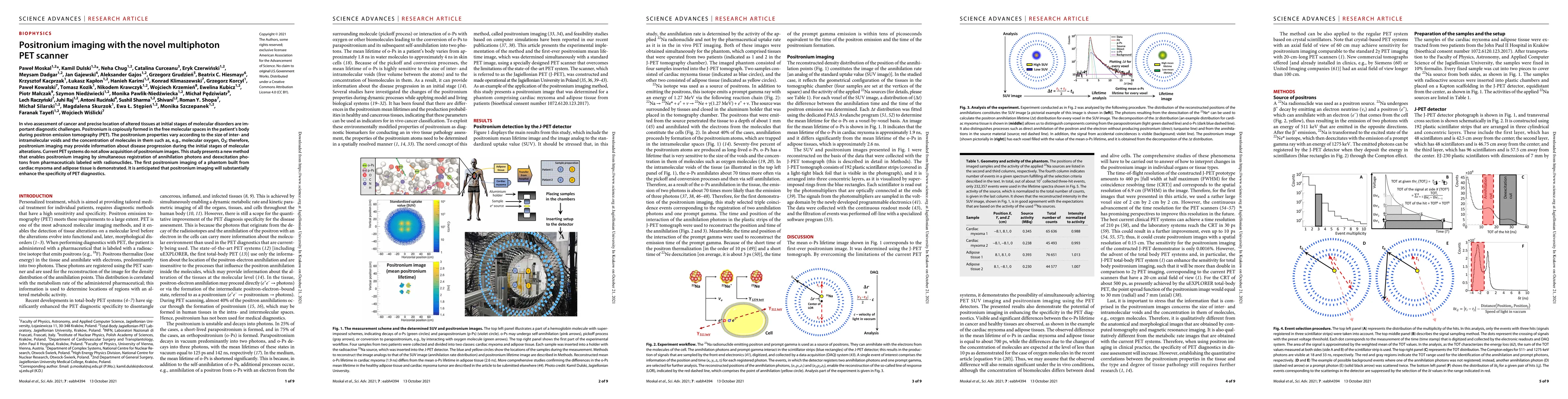
In vivo assessment of cancer and precise location of altered tissues at initial stages of molecular disorders are important diagnostic challenges. Positronium is copiously formed in the free molecular spaces in the patient's body during positron emission tomography (PET). The positronium properties vary according to the size of inter- and intramolecular voids and the concentration of molecules in them such as, e.g., molecular oxygen, O2; therefore, positronium imaging may provide information about disease progression during the initial stages of molecular alterations. Current PET systems do not allow acquisition of positronium images. This study presents a new method that enables positronium imaging by simultaneous registration of annihilation photons and deexcitation photons from pharmaceuticals labeled with radionuclides. The first positronium imaging of a phantom built from cardiac myxoma and adipose tissue is demonstrated. It is anticipated that positronium imaging will substantially enhance the specificity of PET diagnostics.
AI Key Findings
Get AI-generated insights about this paper's methodology, results, significance, and more — seven facets brought into focus.
Impact
Paper Details
Authors
PDF Preview
Key Terms
Citation Network
Current paper (gray), citations (green), references (blue)
Display is limited for performance on very large graphs.
Discussion 0