Pre-examinations Improve Automated Metastases Detection on Cranial MRI
Publication
Metrics
AI Quick Summary
This study investigates the effectiveness of automated metastases detection using cranial MRI. The dual-time approach with contrast-enhanced T1-weighted images (CNNdual_ce) showed high sensitivity and significantly fewer false positives compared to including additional MRI sequences or using only a single time point. Future research should explore alternative detection architectures.
Paper Preview
Abstract
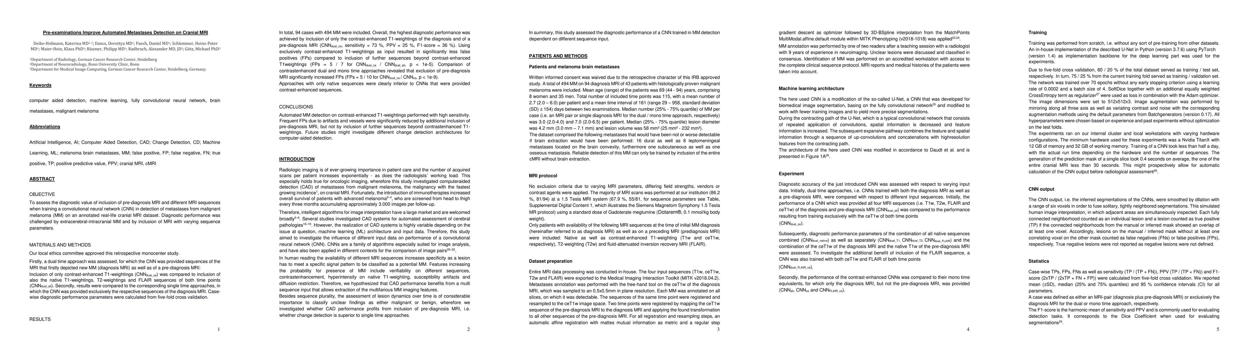
Materials and methods: First, a dual-time approach was assessed, for which the CNN was provided sequences of the MRI that initially depicted new MM (diagnosis MRI) as well as of a prediagnosis MRI: inclusion of only contrast-enhanced T1-weighted images (CNNdual_ce) was compared with inclusion of also the native T1-weighted images, T2-weighted images, and FLAIR sequences of both time points (CNNdual_all).Second, results were compared with the corresponding single time approaches, in which the CNN was provided exclusively the respective sequences of the diagnosis MRI.Casewise diagnostic performance parameters were calculated from 5-fold cross-validation. Results: In total, 94 cases with 494 MMs were included. Overall, the highest diagnostic performance was achieved by inclusion of only the contrast-enhanced T1-weighted images of the diagnosis and of a prediagnosis MRI (CNNdual_ce, sensitivity = 73%, PPV = 25%, F1-score = 36%). Using exclusively contrast-enhanced T1-weighted images as input resulted in significantly less false-positives (FPs) compared with inclusion of further sequences beyond contrast-enhanced T1-weighted images (FPs = 5/7 for CNNdual_ce/CNNdual_all, P < 1e-5). Comparison of contrast-enhanced dual and mono time approaches revealed that exclusion of prediagnosis MRI significantly increased FPs (FPs = 5/10 for CNNdual_ce/CNNce, P < 1e-9).Approaches with only native sequences were clearly inferior to CNNs that were provided contrast-enhanced sequences. Conclusions: Automated MM detection on contrast-enhanced T1-weighted images performed with high sensitivity. Frequent FPs due to artifacts and vessels were significantly reduced by additional inclusion of prediagnosis MRI, but not by inclusion of further sequences beyond contrast-enhanced T1-weighted images. Future studies might investigate different change detection architectures for computer-aided detection.
AI Key Findings
Get AI-generated insights about this paper's methodology, results, significance, and more — seven facets brought into focus.
Impact
Paper Details
Authors
PDF Preview
Key Terms
Citation Network
Current paper (gray), citations (green), references (blue)
Display is limited for performance on very large graphs.
Discussion 0