Predicting Hypoxia in Brain Tumors from Multiparametric MRI
Publication
Metrics
AI Quick Summary
This paper introduces a deep learning model to predict hypoxia in brain tumors using multi-parametric MRI, aiming to replace the expensive and less accessible FMISO PET scans. The study demonstrates strong correlation between predicted and actual FMISO PET signals, indicating MRI's potential for accessible hypoxia detection.
Paper Preview
Abstract
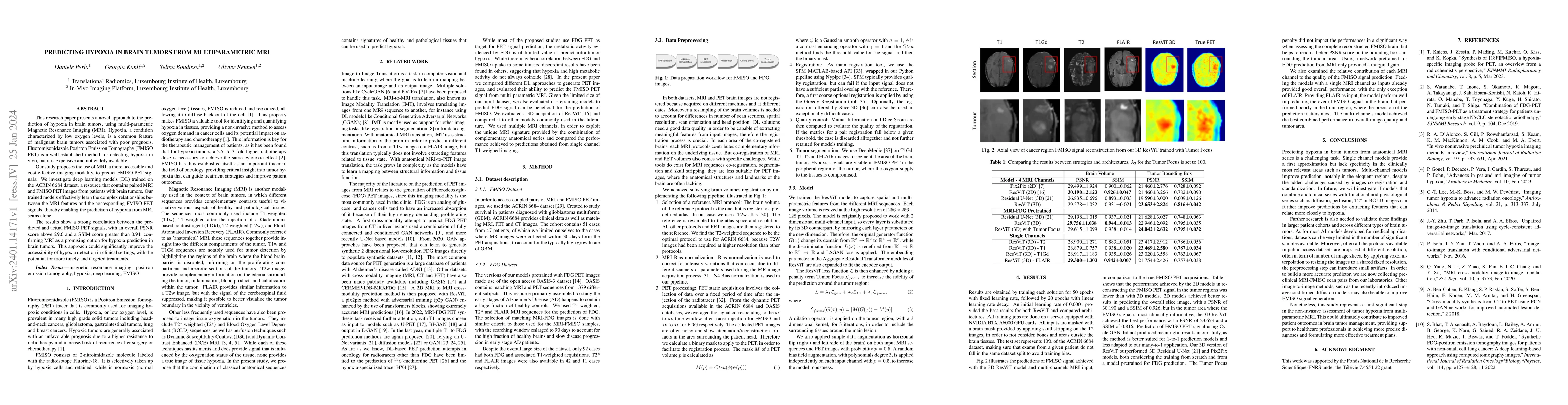
This research paper presents a novel approach to the prediction of hypoxia in brain tumors, using multi-parametric Magnetic Resonance Imaging (MRI). Hypoxia, a condition characterized by low oxygen levels, is a common feature of malignant brain tumors associated with poor prognosis. Fluoromisonidazole Positron Emission Tomography (FMISO PET) is a well-established method for detecting hypoxia in vivo, but it is expensive and not widely available. Our study proposes the use of MRI, a more accessible and cost-effective imaging modality, to predict FMISO PET signals. We investigate deep learning models (DL) trained on the ACRIN 6684 dataset, a resource that contains paired MRI and FMISO PET images from patients with brain tumors. Our trained models effectively learn the complex relationships between the MRI features and the corresponding FMISO PET signals, thereby enabling the prediction of hypoxia from MRI scans alone. The results show a strong correlation between the predicted and actual FMISO PET signals, with an overall PSNR score above 29.6 and a SSIM score greater than 0.94, confirming MRI as a promising option for hypoxia prediction in brain tumors. This approach could significantly improve the accessibility of hypoxia detection in clinical settings, with the potential for more timely and targeted treatments.
AI Key Findings
Get AI-generated insights about this paper's methodology, results, significance, and more — seven facets brought into focus.
Impact
Paper Details
Authors
PDF Preview
Key Terms
Citation Network
Current paper (gray), citations (green), references (blue)
Display is limited for performance on very large graphs.
Discussion 0