Publication
Metrics
AI Quick Summary
This paper develops a deep learning model to predict diabetic macular edema grades from fundus photographs, achieving an ROC-AUC of 0.89, outperforming retinal specialists in specificity and positive predictive value. The model also detects intraretinal and subretinal fluid with high accuracy, demonstrating the potential of deep learning in medical imaging.
Paper Preview
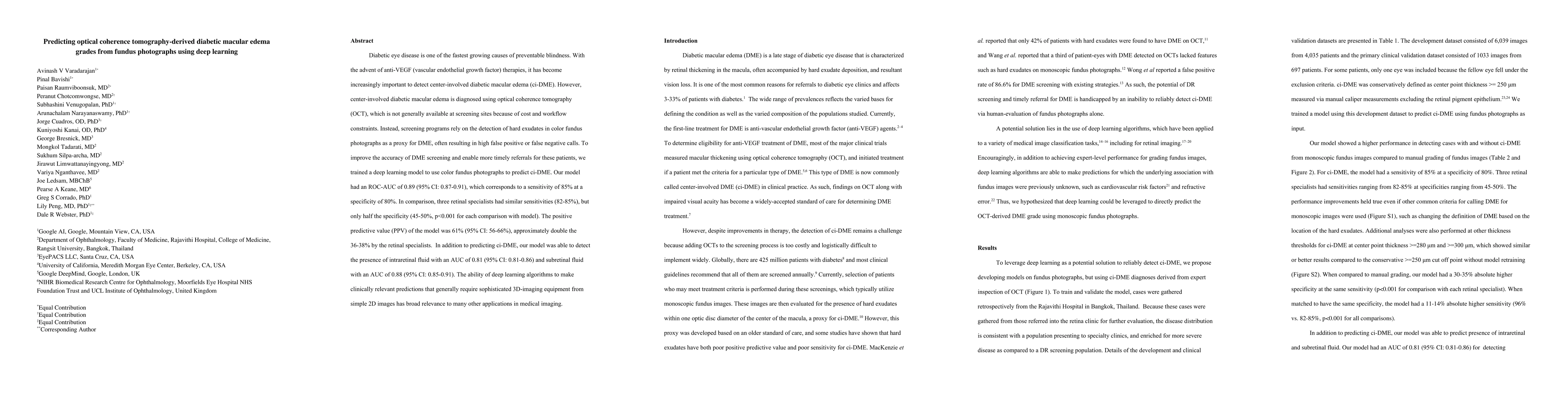
Abstract
Diabetic eye disease is one of the fastest growing causes of preventable blindness. With the advent of anti-VEGF (vascular endothelial growth factor) therapies, it has become increasingly important to detect center-involved diabetic macular edema (ci-DME). However, center-involved diabetic macular edema is diagnosed using optical coherence tomography (OCT), which is not generally available at screening sites because of cost and workflow constraints. Instead, screening programs rely on the detection of hard exudates in color fundus photographs as a proxy for DME, often resulting in high false positive or false negative calls. To improve the accuracy of DME screening, we trained a deep learning model to use color fundus photographs to predict ci-DME. Our model had an ROC-AUC of 0.89 (95% CI: 0.87-0.91), which corresponds to a sensitivity of 85% at a specificity of 80%. In comparison, three retinal specialists had similar sensitivities (82-85%), but only half the specificity (45-50%, p<0.001 for each comparison with model). The positive predictive value (PPV) of the model was 61% (95% CI: 56-66%), approximately double the 36-38% by the retinal specialists. In addition to predicting ci-DME, our model was able to detect the presence of intraretinal fluid with an AUC of 0.81 (95% CI: 0.81-0.86) and subretinal fluid with an AUC of 0.88 (95% CI: 0.85-0.91). The ability of deep learning algorithms to make clinically relevant predictions that generally require sophisticated 3D-imaging equipment from simple 2D images has broad relevance to many other applications in medical imaging.
AI Key Findings
Get AI-generated insights about this paper's methodology, results, significance, and more — seven facets brought into focus.
Impact
Paper Details
PDF Preview
Key Terms
Citation Network
Current paper (gray), citations (green), references (blue)
Display is limited for performance on very large graphs.
Discussion 0