Preoperative brain tumor imaging: models and software for segmentation and standardized reporting
Publication
Metrics
AI Quick Summary
This study develops segmentation models for various brain tumors using AGU-Net architecture, achieving high accuracy in tumor detection. Two software solutions, Raidionics and Raidionics-Slicer, enable automated segmentation and standardized clinical reporting, with segmentation times ranging from 16 to 54 seconds. Future enhancements aim to automate tumor type classification and include post-operative segmentation.
Paper Preview
Abstract
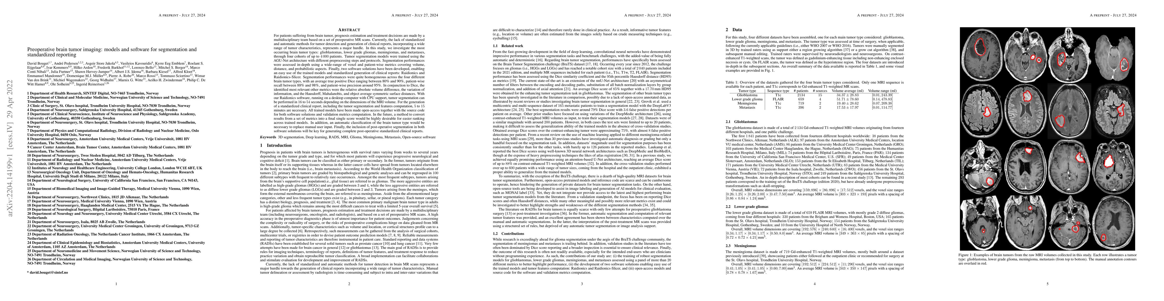
For patients suffering from brain tumor, prognosis estimation and treatment decisions are made by a multidisciplinary team based on a set of preoperative MR scans. Currently, the lack of standardized and automatic methods for tumor detection and generation of clinical reports represents a major hurdle. In this study, we investigate glioblastomas, lower grade gliomas, meningiomas, and metastases, through four cohorts of up to 4000 patients. Tumor segmentation models were trained using the AGU-Net architecture with different preprocessing steps and protocols. Segmentation performances were assessed in-depth using a wide-range of voxel and patient-wise metrics covering volume, distance, and probabilistic aspects. Finally, two software solutions have been developed, enabling an easy use of the trained models and standardized generation of clinical reports: Raidionics and Raidionics-Slicer. Segmentation performances were quite homogeneous across the four different brain tumor types, with an average true positive Dice ranging between 80% and 90%, patient-wise recall between 88% and 98%, and patient-wise precision around 95%. With our Raidionics software, running on a desktop computer with CPU support, tumor segmentation can be performed in 16 to 54 seconds depending on the dimensions of the MRI volume. For the generation of a standardized clinical report, including the tumor segmentation and features computation, 5 to 15 minutes are necessary. All trained models have been made open-access together with the source code for both software solutions and validation metrics computation. In the future, an automatic classification of the brain tumor type would be necessary to replace manual user input. Finally, the inclusion of post-operative segmentation in both software solutions will be key for generating complete post-operative standardized clinical reports.
AI Key Findings
Get AI-generated insights about this paper's methodology, results, significance, and more — seven facets brought into focus.
Impact
Paper Details
Authors
PDF Preview
Key Terms
Citation Network
Current paper (gray), citations (green), references (blue)
Display is limited for performance on very large graphs.
Discussion 0