Publication
Metrics
AI Quick Summary
This paper demonstrates the feasibility of prepolarized MRI (PMRI) for hard biological tissues using a specialized 0.26 T scanner, achieving significant signal-to-noise ratio (SNR) enhancement. The study shows that a 0.5 T prepolarizer module can be activated rapidly, enabling effective imaging of dental and bone samples, with potential for clinical dental imaging to replace X-ray systems.
Paper Preview
Abstract
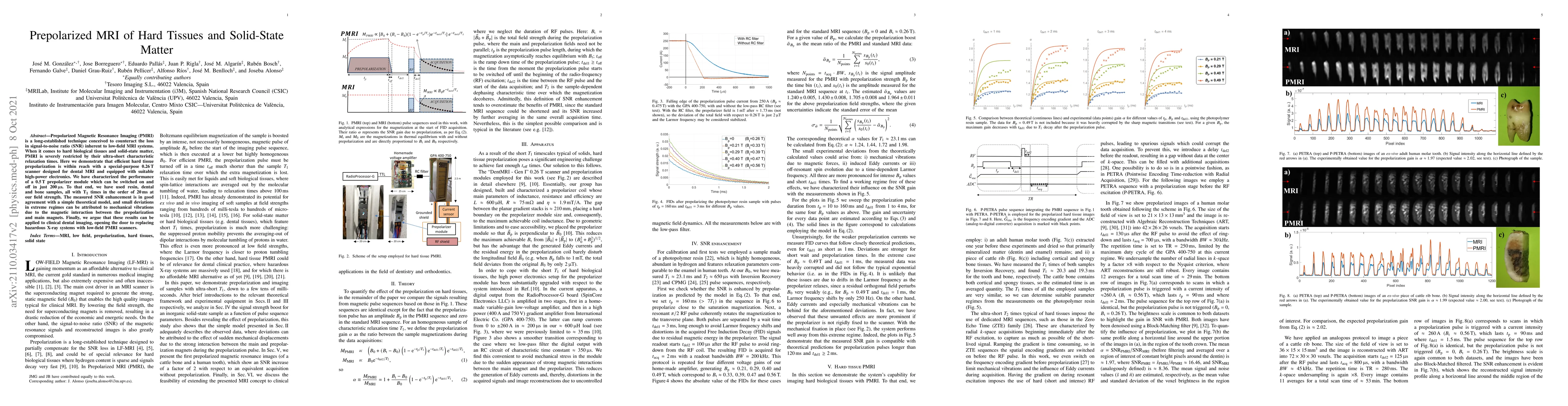
Prepolarized Magnetic Resonance Imaging (PMRI) is a long-established technique conceived to counteract the loss in signal-to-noise ratio (SNR) inherent to low-field MRI systems. When it comes to hard biological tissues and solid-state matter, PMRI is severely restricted by their ultra-short characteristic relaxation times. Here we demonstrate that efficient hard tissue prepolarization is within reach with a special-purpose 0.26 T scanner designed for dental MRI and equipped with suitable high-power electronics. We have characterized the performance of a 0.5 T prepolarizer module which can be switched on and off in just 200 us. To that end, we have used resin, dental and bone samples, all with T1 times in the order of 20 ms at our field strength. The measured SNR enhancement is in good agreement with a simple theoretical model, and small deviations in extreme regimes can be attributed to mechanical vibrations due to the magnetic interaction between the prepolarization and main magnets. Finally, we argue that these results can be applied to clinical dental imaging, opening the door to replacing hazardous X-ray systems with low-field PMRI scanners.
AI Key Findings
Get AI-generated insights about this paper's methodology, results, significance, and more — seven facets brought into focus.
Impact
Paper Details
Authors
PDF Preview
Key Terms
Citation Network
Current paper (gray), citations (green), references (blue)
Display is limited for performance on very large graphs.
Discussion 0