Publication
Metrics
AI Quick Summary
This paper proposes a novel preprocessing technique to automate the early detection of cervical cancer by eliminating specular reflections from colposcopic images and identifying the region of interest in the cervix, thereby facilitating accurate segmentation and analysis.
Paper Preview
Abstract
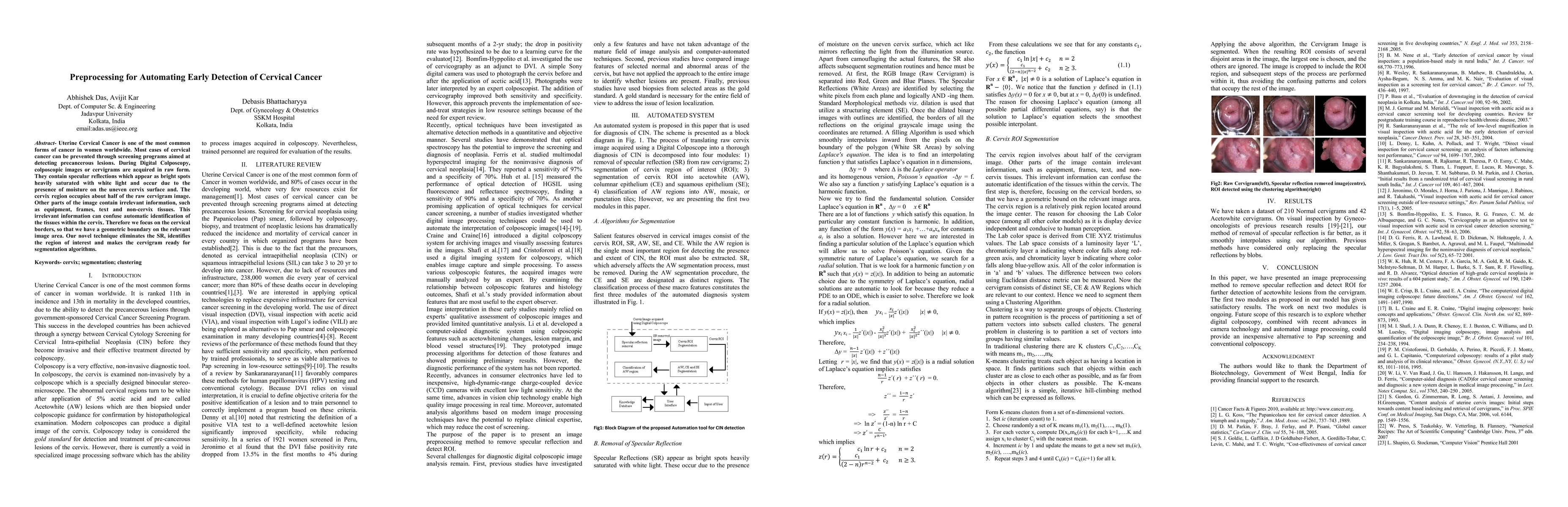
Uterine Cervical Cancer is one of the most common forms of cancer in women worldwide. Most cases of cervical cancer can be prevented through screening programs aimed at detecting precancerous lesions. During Digital Colposcopy, colposcopic images or cervigrams are acquired in raw form. They contain specular reflections which appear as bright spots heavily saturated with white light and occur due to the presence of moisture on the uneven cervix surface and. The cervix region occupies about half of the raw cervigram image. Other parts of the image contain irrelevant information, such as equipment, frames, text and non-cervix tissues. This irrelevant information can confuse automatic identification of the tissues within the cervix. Therefore we focus on the cervical borders, so that we have a geometric boundary on the relevant image area. Our novel technique eliminates the SR, identifies the region of interest and makes the cervigram ready for segmentation algorithms.
AI Key Findings
Get AI-generated insights about this paper's methodology, results, significance, and more — seven facets brought into focus.
Impact
Paper Details
PDF Preview
Key Terms
Citation Network
Current paper (gray), citations (green), references (blue)
Display is limited for performance on very large graphs.
Discussion 0