Publication
Metrics
AI Quick Summary
POSSIBLE microscopy introduces a method for ultra-superresolution imaging by selectively localizing the brightest molecules, thereby filtering out less precise localizations and achieving higher resolution images. This technique demonstrates improved resolution and clarity in imaging molecular clusters in NIH3T3 fibroblast cells compared to traditional SMLM methods.
Paper Preview
Abstract
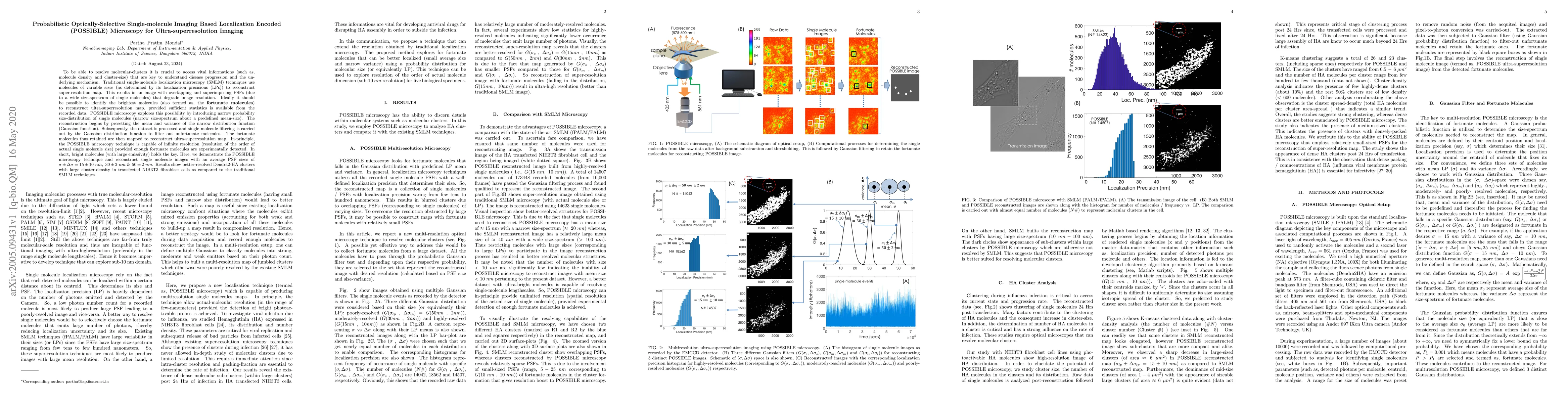
To be able to resolve molecular-clusters it is crucial to access vital informations (such as, molecule density and cluster-size) that are key to understand disease progression and the underlying mechanism. Traditional single-molecule localization microscopy (SMLM) techniques use molecules of variable sizes (as determined by its localization precisions (LPs)) to reconstruct super-resolution map. This results in an image with overlapping and superimposing PSFs (due to a wide size-spectrum of single molecules) that degrade image resolution. Ideally it should be possible to identify the brightest molecules (also termed as, fortunate molecules) to reconstruct ultra-superresolution map, provided sufficient statistics is available from the recorded data. POSSIBLE microscopy explores this possibility by introducing narrow probability size-distribution of single molecules (narrow size-spectrum about a predefined mean-size). The reconstruction begins by presetting the mean and variance of the narrow distribution function (Gaussian function). Subsequently, the dataset is processed and single molecule filtering is carried out by the Gaussian distribution function to filter out unfortunate molecules. The fortunate molecules thus retained are then mapped to reconstruct ultra-superresolution map. In-principle, the POSSIBLE microscopy technique is capable of infinite resolution (resolution of the order of actual single molecule size) provided enough fortunate molecules are experimentally detected. In short, bright molecules (with large emissivity) holds the key. Here, we demonstrate the POSSIBLE microscopy technique and reconstruct single molecule images with an average PSF sizes of 15 nm, 30 nm and 50 nm. Results show better-resolved Dendra2-HA clusters with large cluster-density in transfected NIH3T3 fibroblast cells as compared to the traditional SMLM techniques.
AI Key Findings
Get AI-generated insights about this paper's methodology, results, significance, and more — seven facets brought into focus.
Impact
Paper Details
Authors
PDF Preview
Key Terms
Citation Network
Current paper (gray), citations (green), references (blue)
Display is limited for performance on very large graphs.
Discussion 0