Probing chirality with inelastic electron-light scattering
Publication
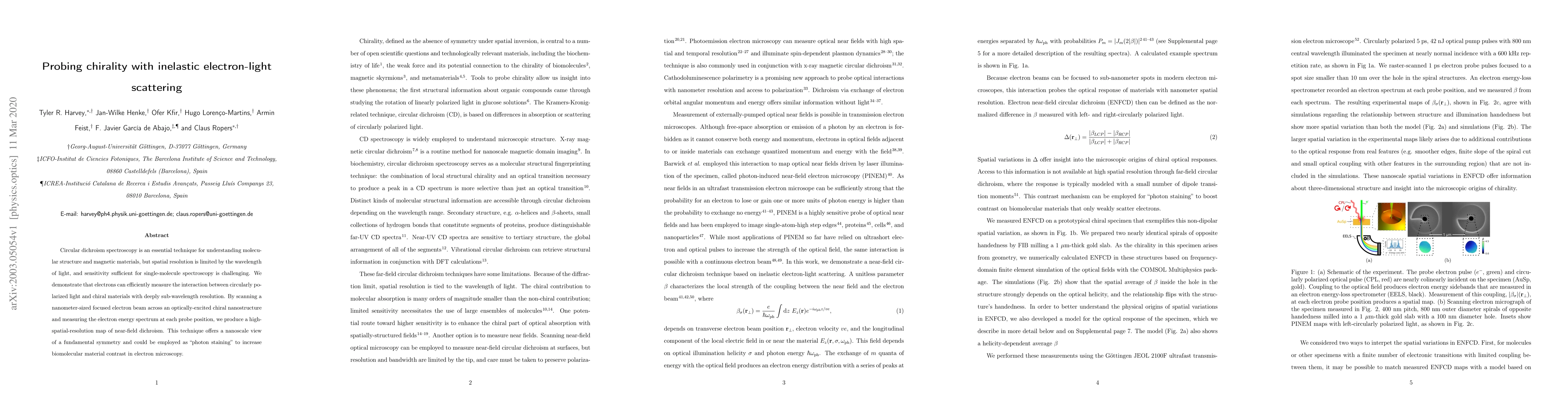
Metrics
AI Quick Summary
This research introduces a novel method using electrons to achieve deeply sub-wavelength resolution in probing chirality of materials, overcoming limitations of traditional circular dichroism spectroscopy. The technique, called 'photon staining', maps near-field dichroism, potentially enhancing contrast in electron microscopy for biomolecular materials.
Paper Preview
Abstract
Circular dichroism spectroscopy is an essential technique for understanding molecular structure and magnetic materials, but spatial resolution is limited by the wavelength of light, and sensitivity sufficient for single-molecule spectroscopy is challenging. We demonstrate that electrons can efficiently measure the interaction between circularly polarized light and chiral materials with deeply sub-wavelength resolution. By scanning a nanometer-sized focused electron beam across an optically-excited chiral nanostructure and measuring the electron energy spectrum at each probe position, we produce a high-spatial-resolution map of near-field dichroism. This technique offers a nanoscale view of a fundamental symmetry and could be employed as "photon staining" to increase biomolecular material contrast in electron microscopy.
AI Key Findings
Get AI-generated insights about this paper's methodology, results, significance, and more — seven facets brought into focus.
Impact
Paper Details
Authors
PDF Preview
Key Terms
Citation Network
Current paper (gray), citations (green), references (blue)
Display is limited for performance on very large graphs.
Discussion 0