Publication
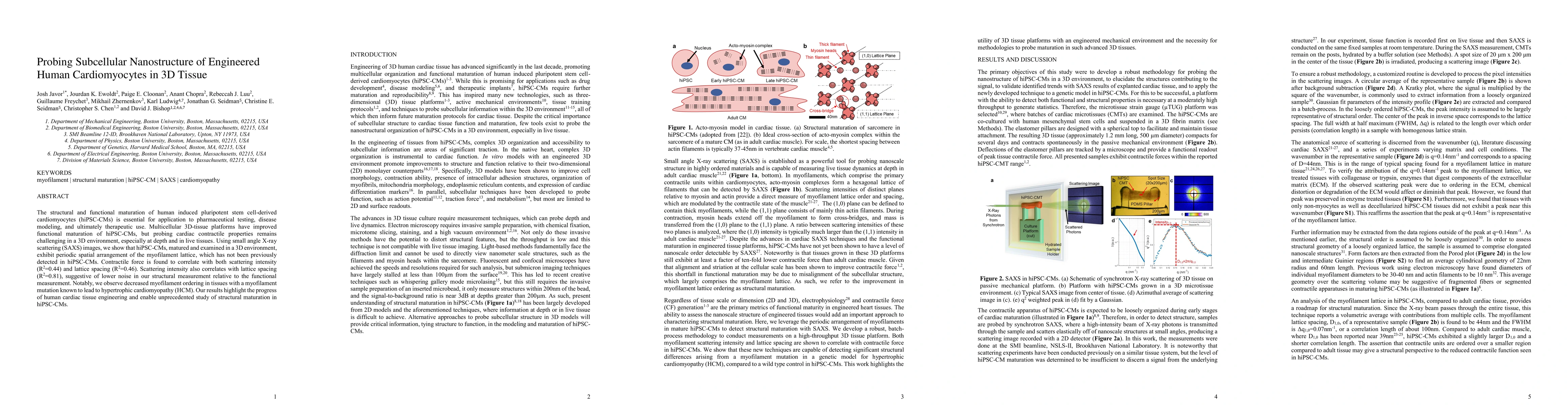
Metrics
AI Quick Summary
This study uses small angle X-ray scattering to reveal the periodic spatial arrangement of myofilament lattice in 3D tissue-engineered human iPSC-CMs, correlating contractile force with scattering intensity and lattice spacing. It also identifies decreased myofilament ordering in HCM-linked mutations, underscoring the potential of 3D tissue platforms for cardiac tissue engineering.
Paper Preview
Abstract
The structural and functional maturation of human induced pluripotent stem cell-derived cardiomyocytes (hiPSC-CMs) is essential for application to pharmaceutical testing, disease modeling, and ultimately therapeutic use. Multicellular 3D-tissue platforms have improved functional maturation of hiPSC-CMs, but probing cardiac contractile properties remains challenging in a 3D environment, especially at depth and in live tissues. Using small angle X-ray scattering (SAXS) images, we show that hiPSC-CMs, matured and examined in a 3D environment, exhibit periodic spatial arrangement of the myofilament lattice, which has not been previously detected in hiPSC-CMs. Contractile force is found to correlate with both scattering intensity (R2=0.44) and lattice spacing (R2=0.46). Scattering intensity also correlates with lattice spacing (R2=0.81), suggestive of lower noise in our structural measurement relative to the functional measurement. Notably, we observe decreased myofilament ordering in tissues with a myofilament mutation known to lead to hypertrophic cardiomyopathy (HCM). Our results highlight the progress of human cardiac tissue engineering and enable unprecedented study of structural maturation in hiPSC-CMs.
AI Key Findings
Get AI-generated insights about this paper's methodology, results, significance, and more — seven facets brought into focus.
Impact
Paper Details
Authors
PDF Preview
Key Terms
Citation Network
Current paper (gray), citations (green), references (blue)
Display is limited for performance on very large graphs.
Discussion 0