01
MethodologyHow they did it
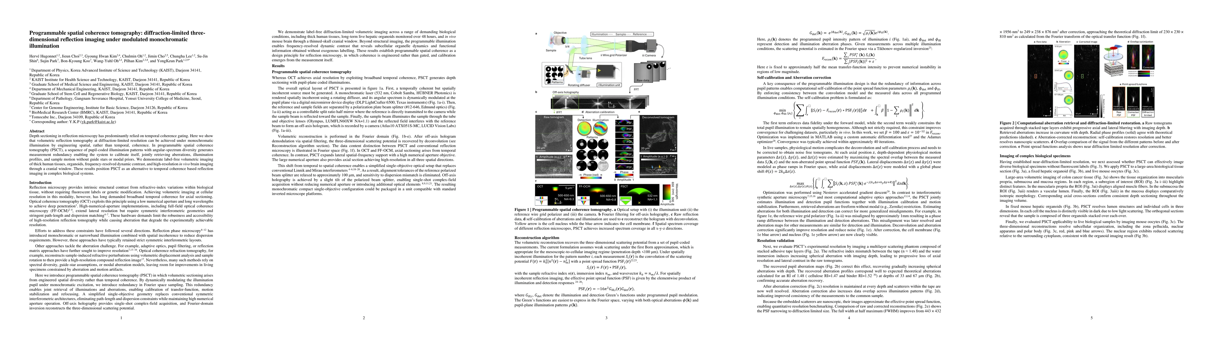
PSCT uses a monochromatic, spatially incoherent light source whose angular spectrum is dynamically modulated at the pupil plane via a DLP to create a sequence of pupil-coded illumination patterns. Off-axis holography with a single-objective, non-interferometric path-length setup records complex-field measurements, which are then reconstructed in the Fourier domain to yield a three-dimensional scattering potential. The approach jointly estimates illuminations, aberrations, and sample motion by leveraging redundancy in Fourier space to calibrate transfer-functions and enable refocusing and motion stabilization without guide stars or modal priors.
Discussion 0