01
MethodologyHow they did it
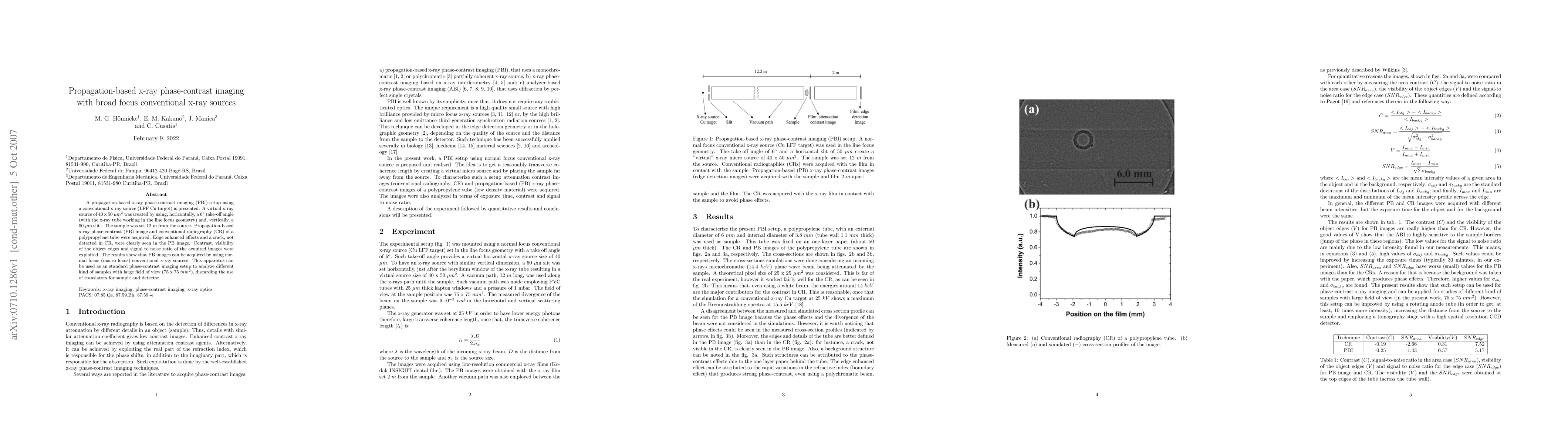
The research presents a propagation-based x-ray phase-contrast imaging (PBI) setup using a conventional x-ray source (LFF Cu target) in line focus geometry. A virtual microfocus x-ray source of 40 x 50 $\mu$$m^2$ is created with a 6$^\circ$ take-off angle horizontally and a 50 $\mu$m slit vertically. Samples are placed 12 m from the source, and both propagation-based x-ray phase-contrast (PB) images and conventional radiography (CR) are acquired.
Discussion 0